Tutorial: Intro to VRmol
Tutorial: Intro to VRmolIntroductionVR Mode Getting Started VR DeviceEntering VR Scene (V)VR MenuLoading Structure (V)Main StructureLigandSurfaceColor SchemeMeasurement (V)Drag (V)Fragmentation (V)Structure Editing (V)Genomic Variations (V)Drug Docking (V)Speech Recognition (V)NonVR Mode Getting Started Mouse UsageLoading Structure Main StructureLigandSurfaceColor SchemeFragmentation (V)Structure Editing (V)Genomic Variations (V)Drug Docking (V)Sharing by URLURL OptionsExamples for API VRmol API ServicesGenomic VariationsSequence ConservationDrug data APIEMDBAutoDockSpeech RecognitionContacts
Introduction
Welcome to the VRmol tutorial. VRmol is an easy-to-use web-based tool capable of visualizing and integratively analyzing biomolecular structures in Virtual Reality environment. It connects to multiple resources, such as disease and drug-related databases, enabling it to serve as an integrative platform for structure-based translational researches and drug design. It can be freely accessed online by typing https://VRmol.net in local browser. The following pages present a brief self-guided tour of the VRmol.
The tutorial includes three sections:
- VR Mode will guide you to experience VRmol in Virtual Reality environment.
- NonVR Mode will guide you to experience VRmol on the NonVR.
- Sharing URL will guide you to use VRmol with a sharing URL which can be embedded into your own webpage with only a single line HTML code.
VR Mode
Getting Started
VR Device
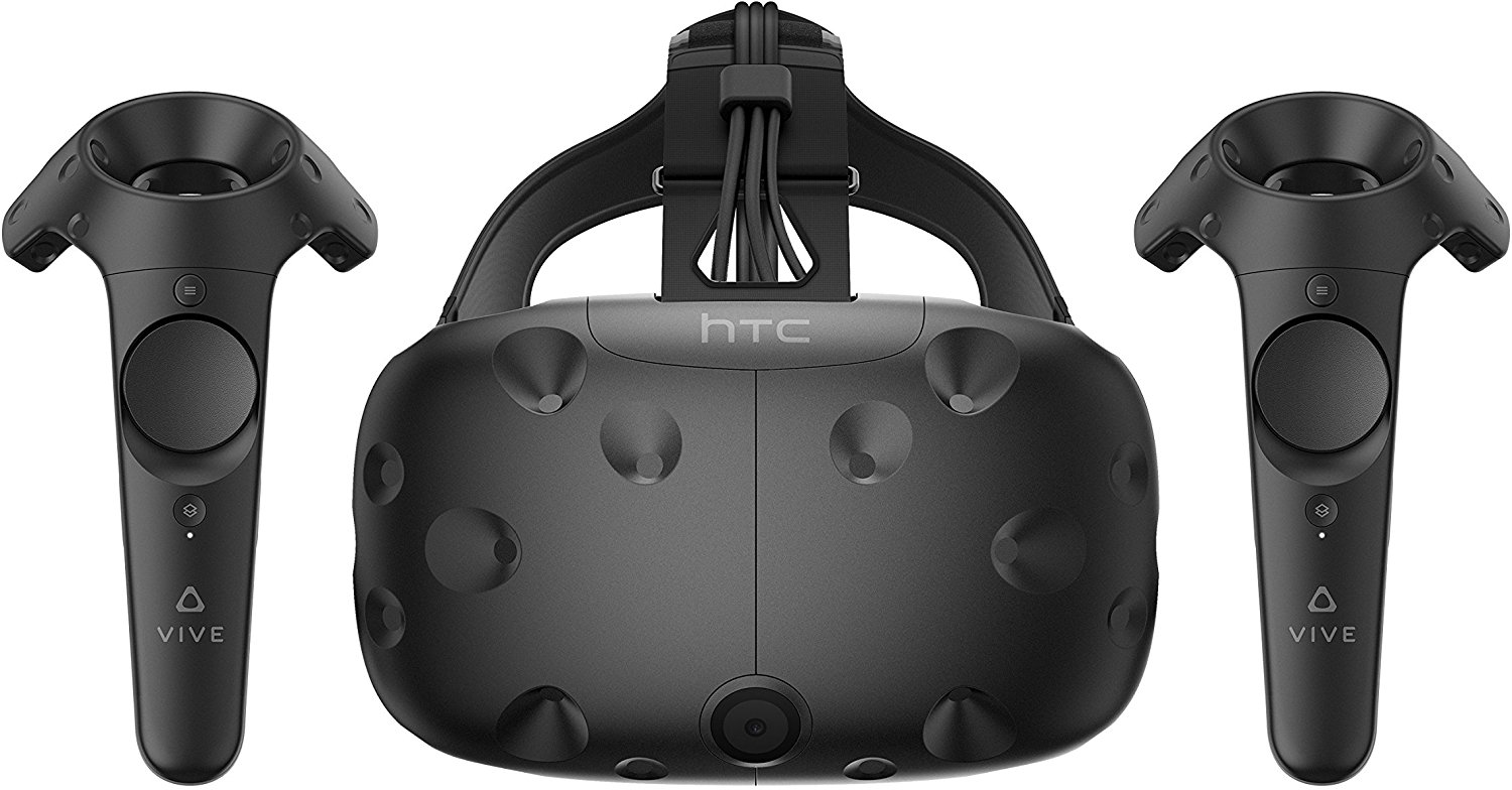
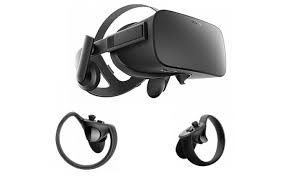
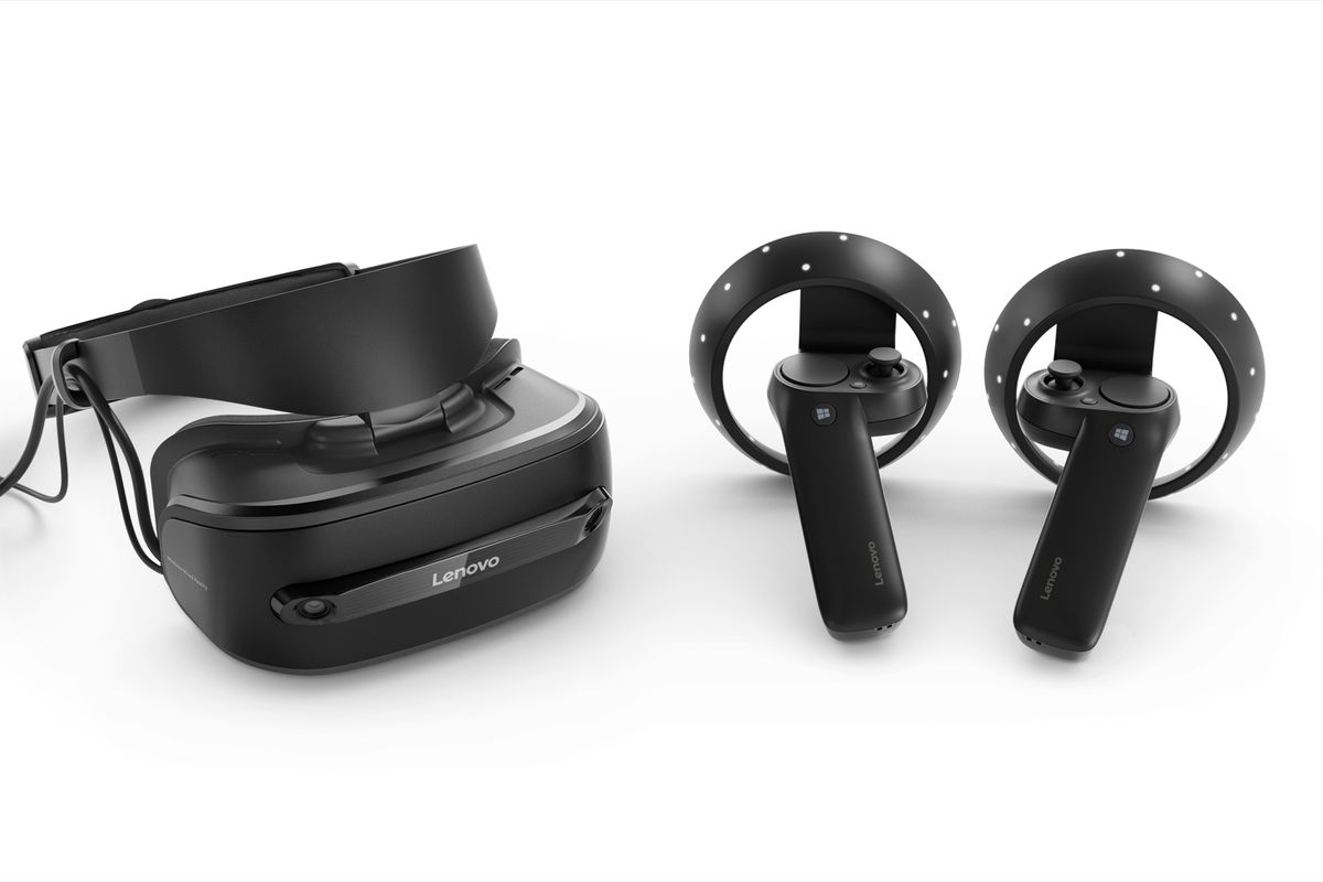
For experiencing VR Scene in VRmol, a VR device is required. VR devices such as HTC Vive, Oculus Rift, and Microsoft Mixed Reality are well-supported by VRmol. Specifically, for HTC Vive, you should install SteamVR from Steam library and setup the room environment in advance. The compatibility between these VR devices and web browsers listed in the following table has been successfully tested in VRmol.
| Device | OS | Web Browser |
|---|---|---|
| Windows Mixed Reality* | Windows 10 (build 1903+) | Microsoft Edge(v81+), Google Chrome(v81+), Firefox Reality |
| HTC VIVE | Windows 10 (build 1903+) | Microsoft Edge(v81+), Google Chrome(v81+), Firefox Reality |
| Oculus Rift | Windows 10 (build 1903+) | Microsoft Edge(v81+), Google Chrome(v81+), Firefox Reality |
*: Recommanded.
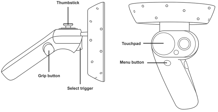
The operating elements in the VR device and their functions in VR mode are described as follows:
- Trigger. Select an object or enter a function by pressing TriggerButton.
- ThumbStickButton &ThumbPadButton for Structure translation and rotation. For HTC Vive, Up and Down area in ThumbStickButton is used to transit the structure. Left and Right area in ThumbStickButton is used to rotate the structure. The axis can be changed in VR menu. For Oculus Rift and Windows Mixed Reality, Up and Down area in ThumbPad is used to transit the structure. Left and Right area in ThumbPad is used to rotate the structure. The axis can be changed in VR menu.
- Grip Button. Show VR menu.
VR Controllers input map is shown below:
| HTC Vive | Oculus Rift | Windows Mixed Reality |
|---|---|---|
 |  |  |
 |  |  |
| Setup docs | Setup docs | Setup docs, Hardware guidelines |
Notably, the mouse in the desktop interface can also control structural operations in VR environment.
Entering VR Scene (V)
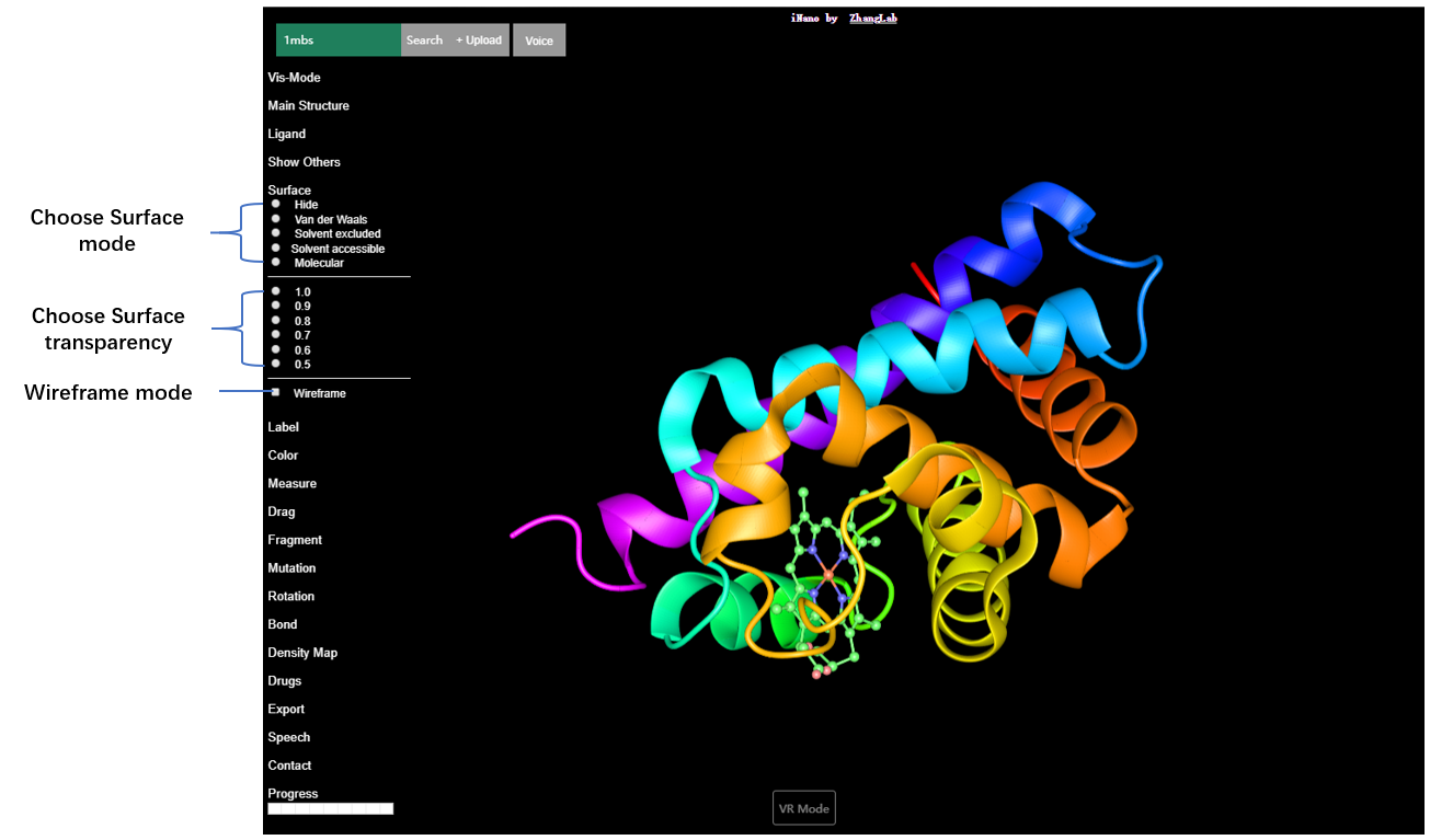
Access VRmol at https://VRmol.net through a WebXR enabled web browser, such as Google Chrome(v81+), Microsoft Edge(v81+) and Firefox Reality. Once you finish typing the website and press Enter button, an VRmol user interface will appear as follows: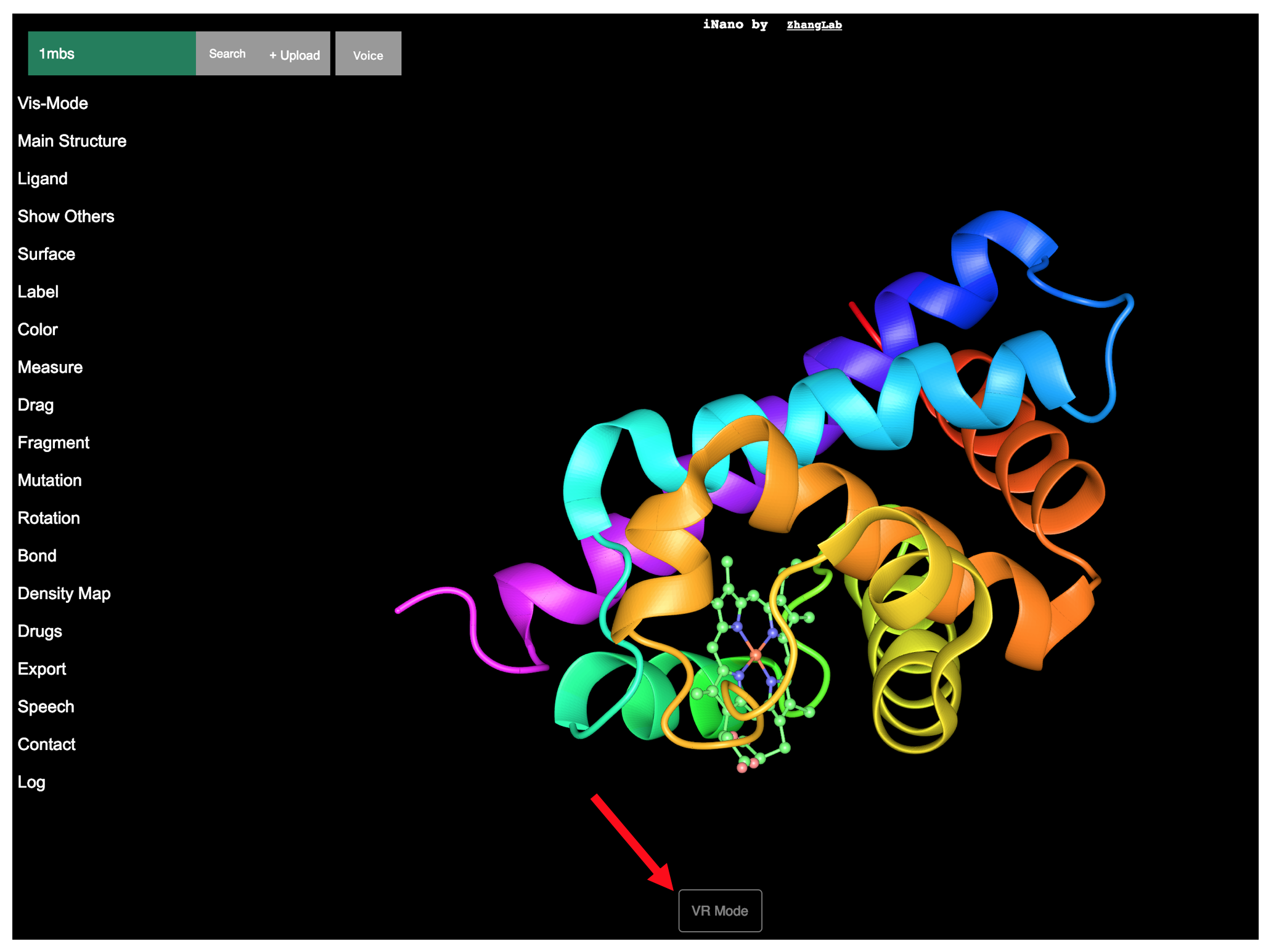
Click VR Mode button at the bottom of the VRmol user interface using the left mouse button, and the VR starting interface will appear as follows. Click the ENTER VR button by using the left mouse button to enter VR Scene.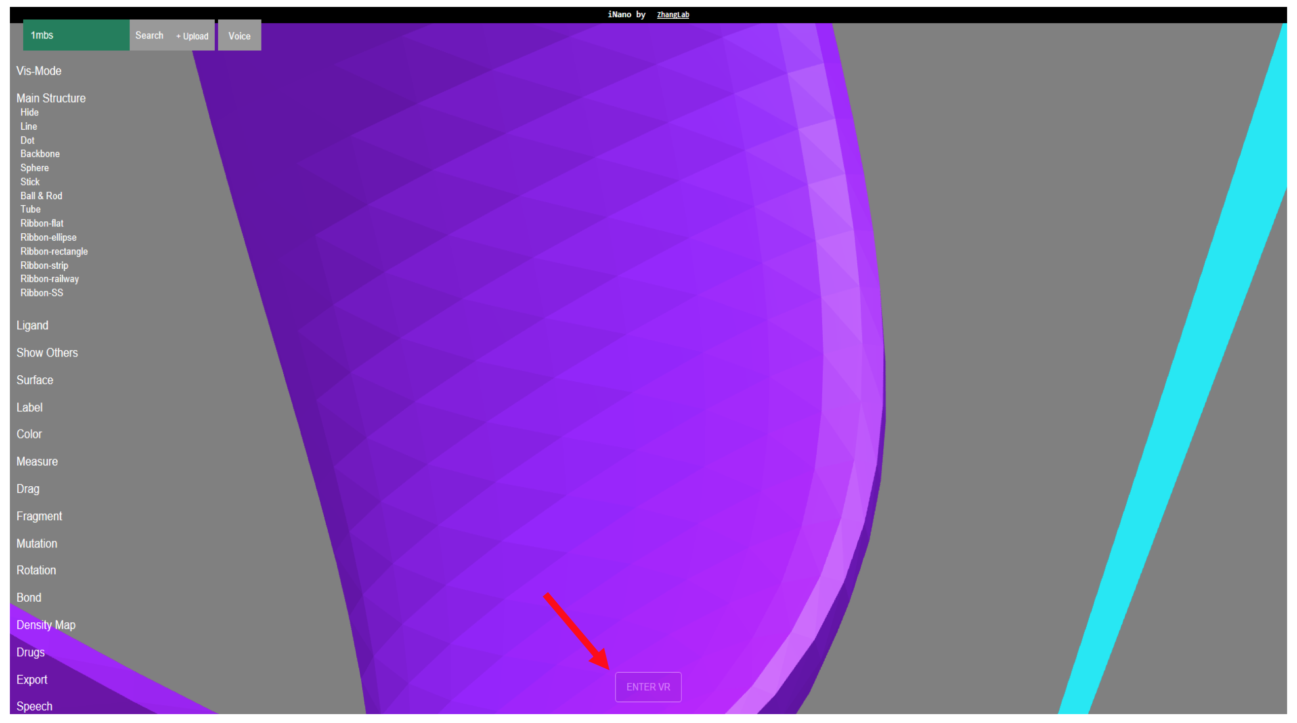
Alternatively, you can also point the laser tip of the controller to ENTER VR button and press the trigger to Enter VR Scene.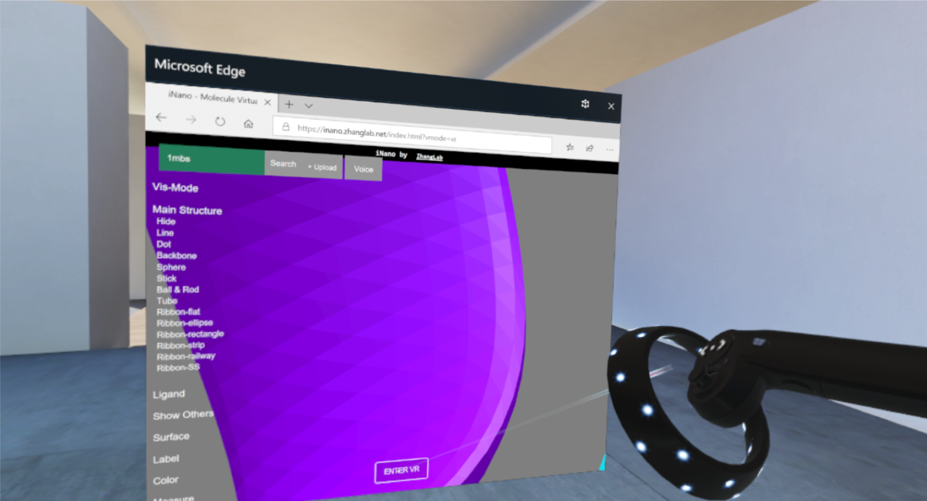
Here is an example video of entering VR Mode.
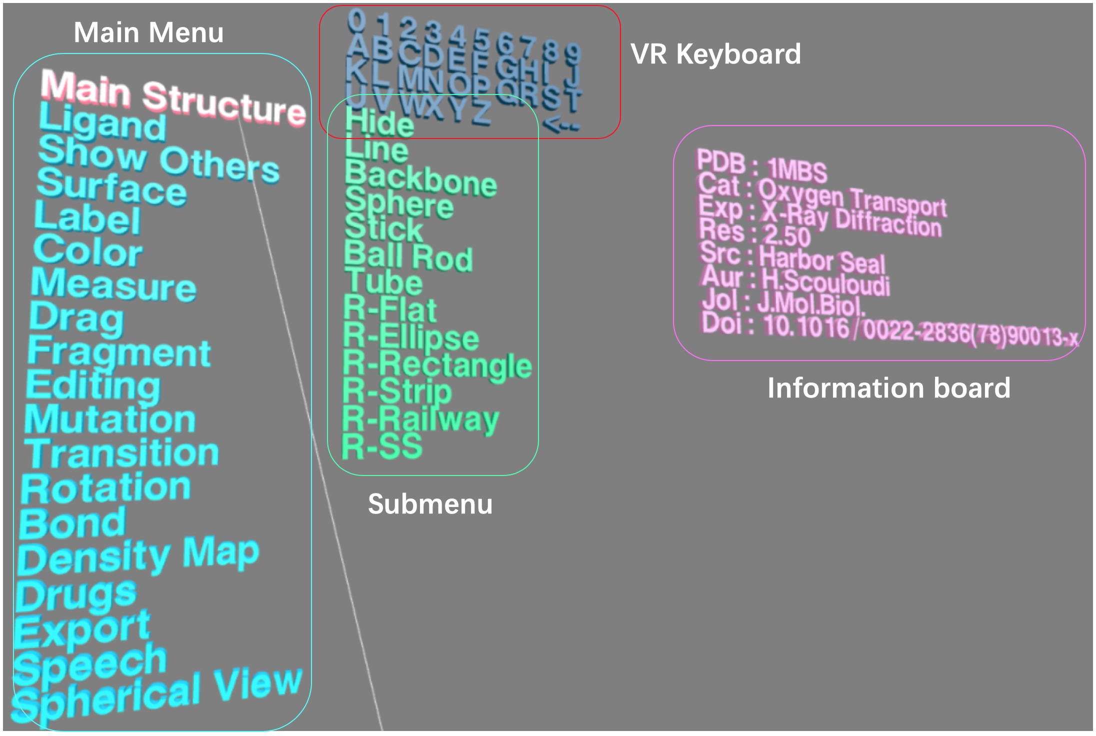
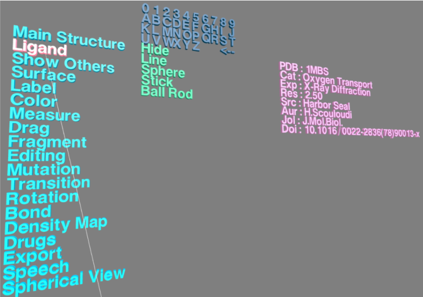
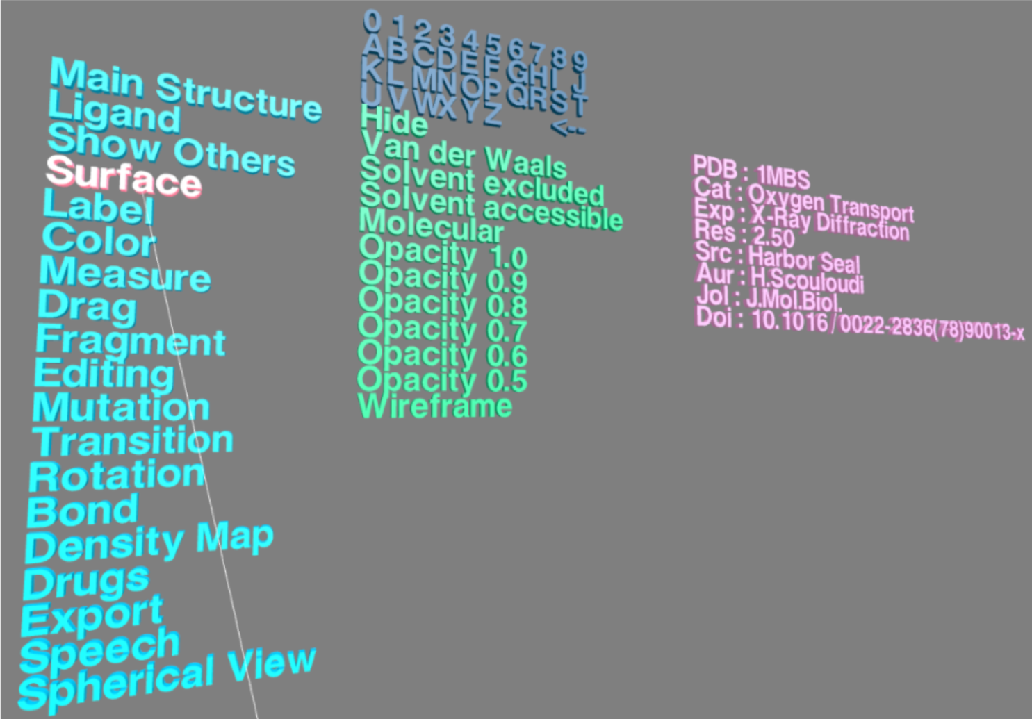
VR Menu
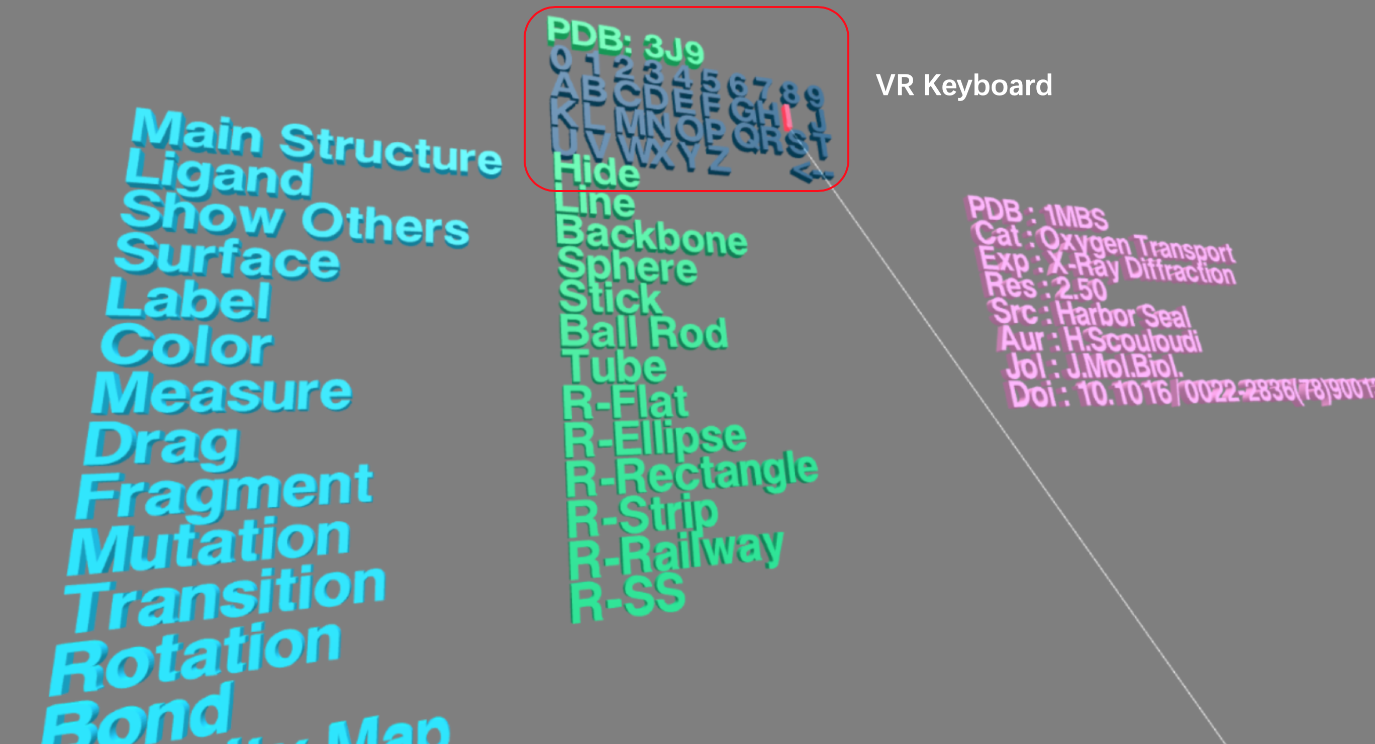
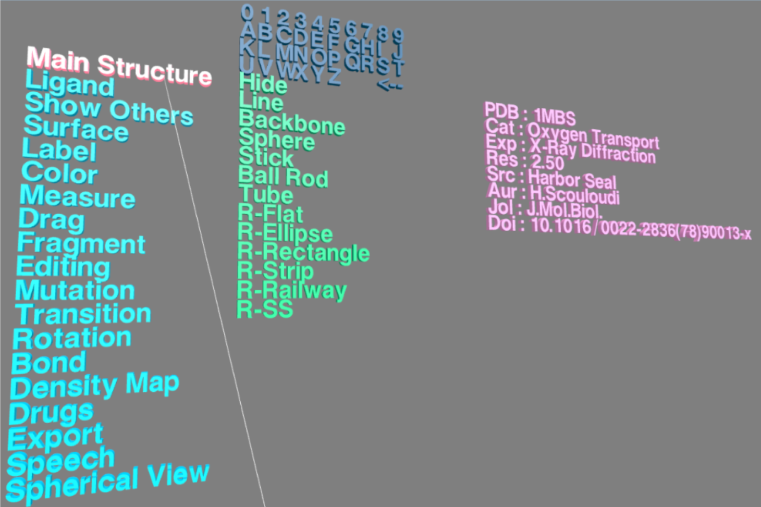
Press Menu button on the controller, a VR menu will appear in front of you in VR Scene (as shown below).The VR menu contains four parts. The top part (labeled in red box) is a VR keyboard including a 26-character alphabet, 10 numbers and a deletion (<--) button; the other three parts consist of a main menu colored in blue, a submenu in green and an information board in pink.
Loading Structure (V)
Press Menu button on the controller to get VR menu. By pointing the laser tip of the controller to alphabet or number in the VR keyboard and pressing the trigger, and a 4-character PDB ID can be typed and the corresponding structure will be afterwards loaded into the VR Scene. Alternatively, the structure in PDB format can also be loaded into the VR environment by typing its PDB ID, inputing a remote URL (such as https://files.rcsb.org/view/1mbs.pdb) or uploading a local structural file in the VRmol Desktop user interface.
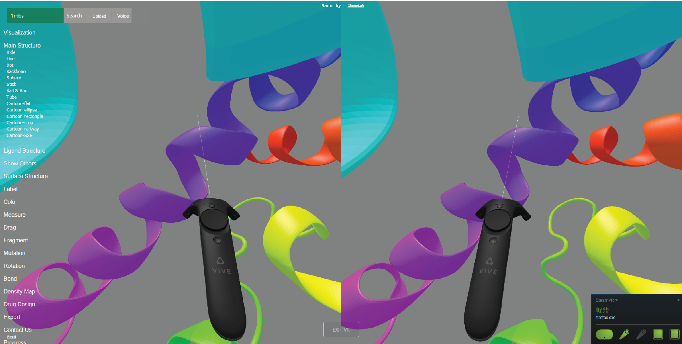
A screenshot of structure visualization with stereo view.
Press Menu button on the controller again, you will go back to the VR menu. By pointing the laser tip to options in other three parts of the menu and pressing the trigger, you can experience structrual visualization styles and analysis in VR Scene, as is concretely described in the following sections.
Here is a video for eploring the structure in VRmol VR mode.
Main Structure
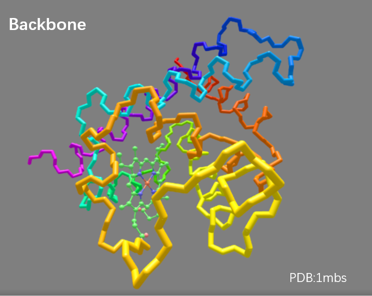
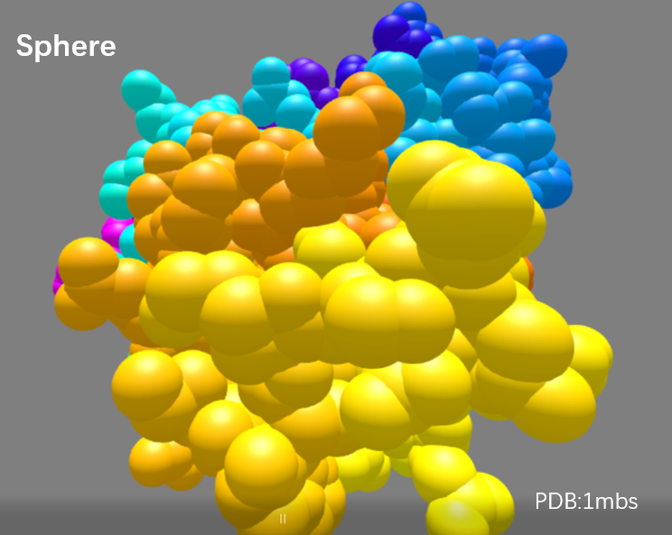
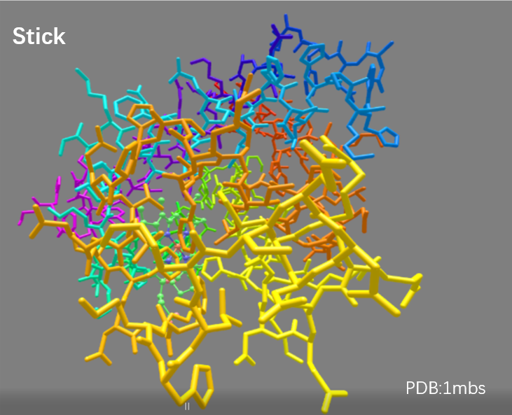
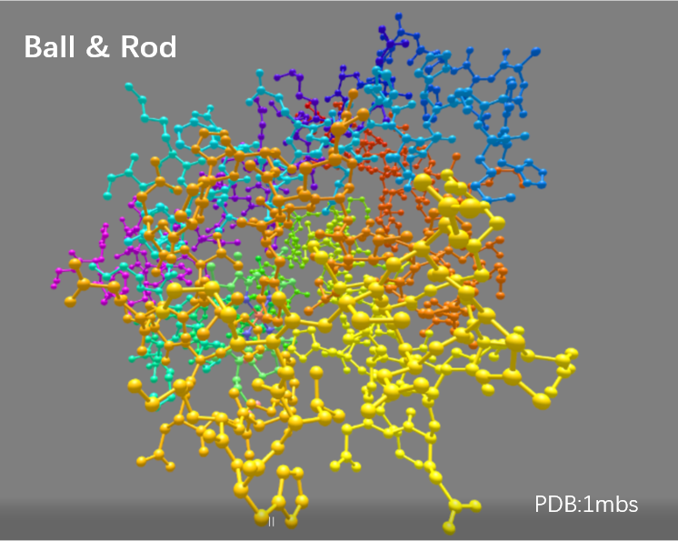
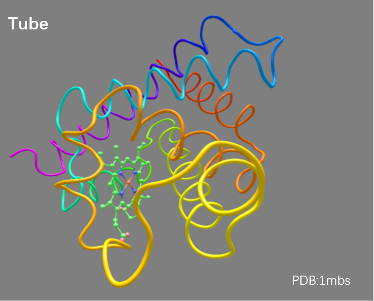
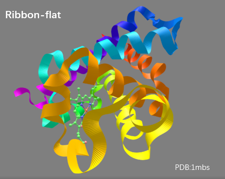
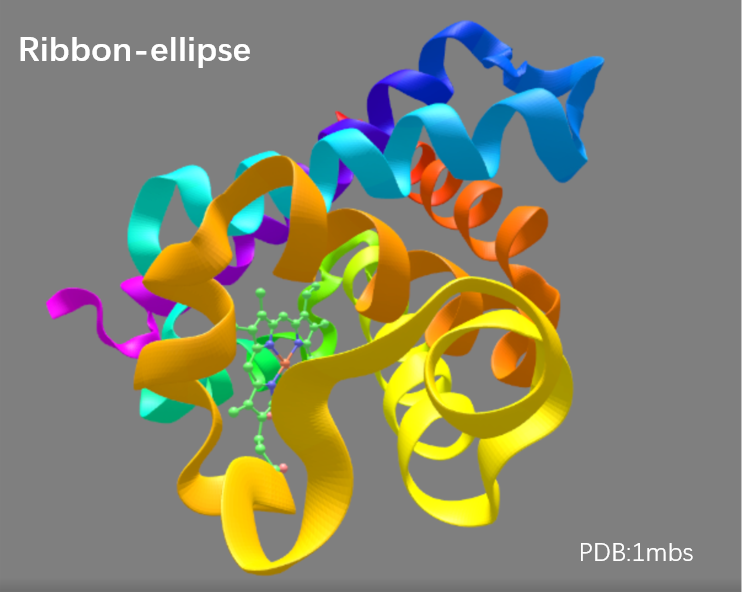
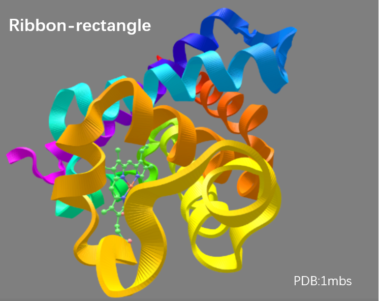
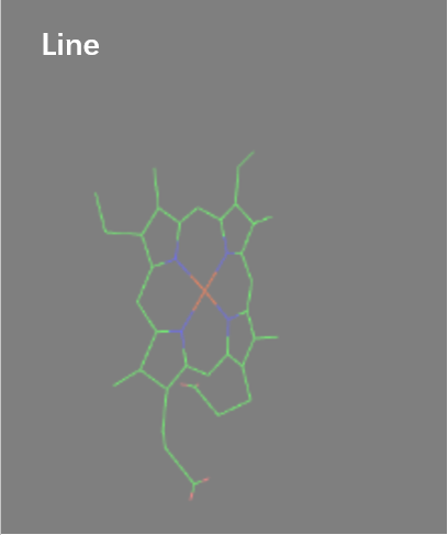
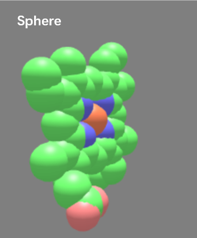
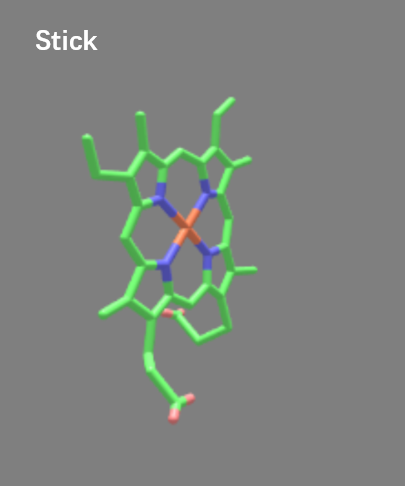
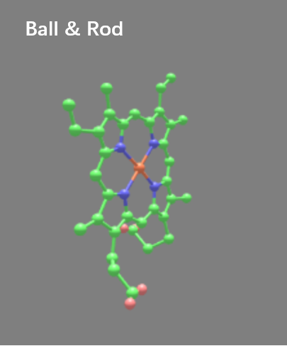
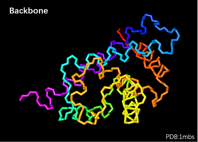
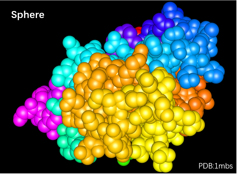
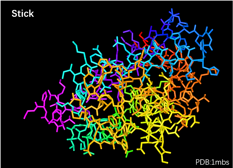
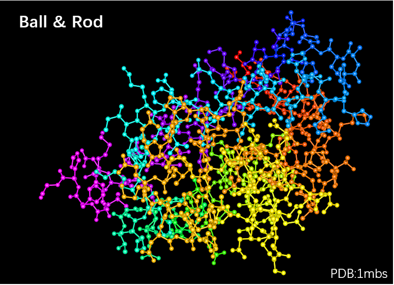
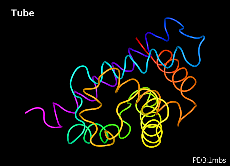
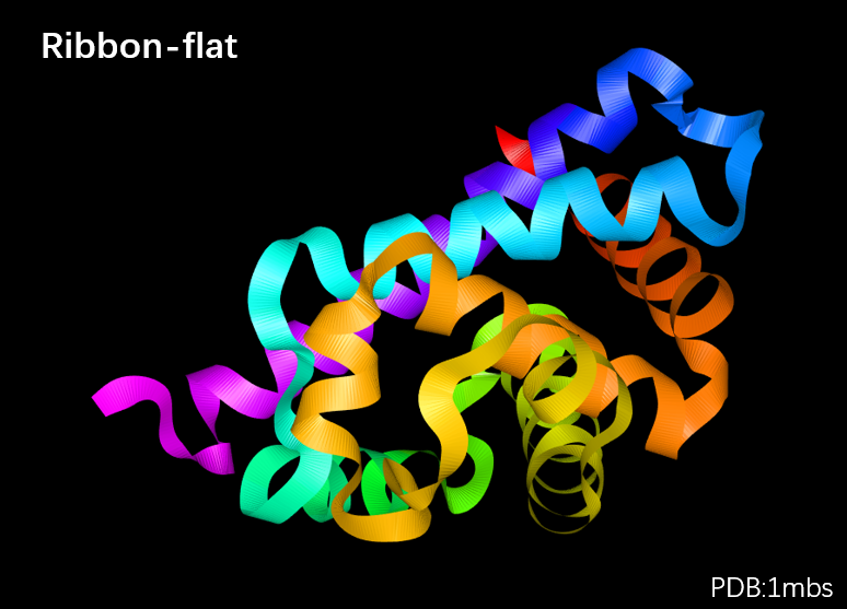
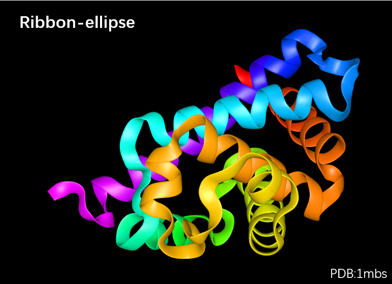
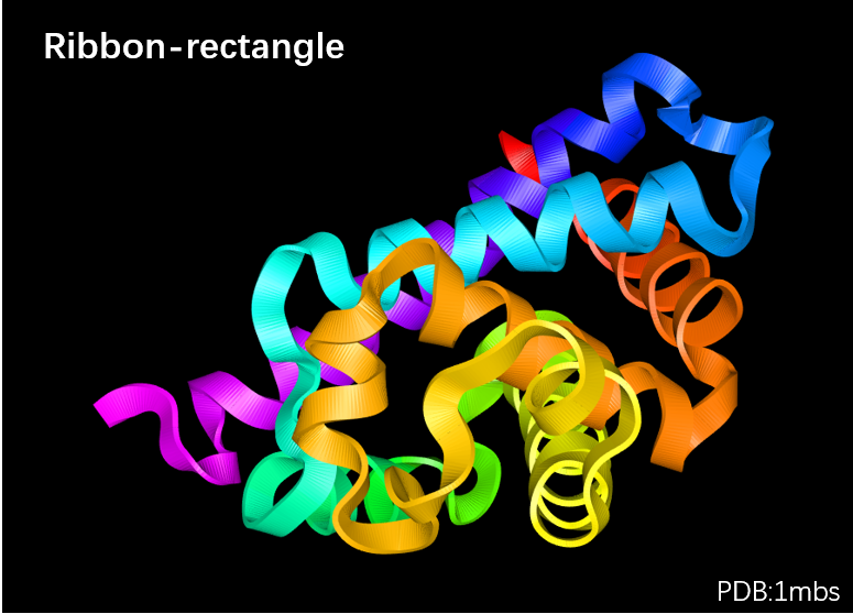
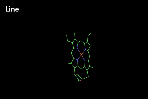
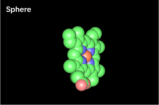
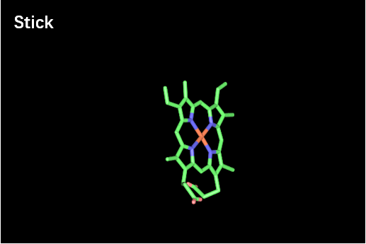
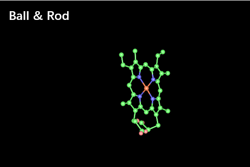
Point the laser tip to "Main Structure" option in VR menu and press the trigger, and 12 different representation styles will appear in the submenu board, including Line, Backbone, Sphere, Stick, Ball & Rod, Tube, Ribbon-flat, Ribbon-ellipse, Ribbon-rectangle, Ribbon-strip, Ribbon-railway, Ribbon-SS. You can visualize the input structure in your favorable style by choosing the specific representation style.
Menu → Main Structure → Line
Menu → Main Structure → Backbone
Menu → Main Structure → Sphere
Menu → Main Structure → Stick
Menu → Main Structure → Ball & Rod
Menu → Main Structure → Tube
Menu → Main Structure → Ribbon-flat
Menu → Main Structure → Ribbon-ellipse
Menu → Main Structure → Ribbon-rectangle
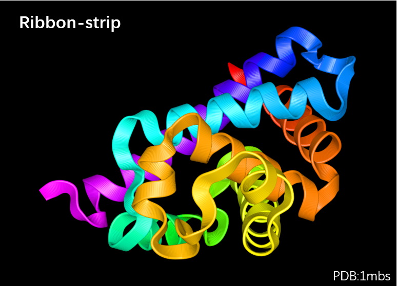
Menu → Main Structure → Ribbon-strip
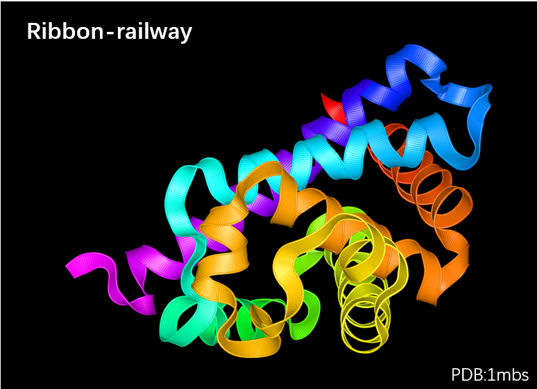
Menu → Main Structure → Ribbon-railway
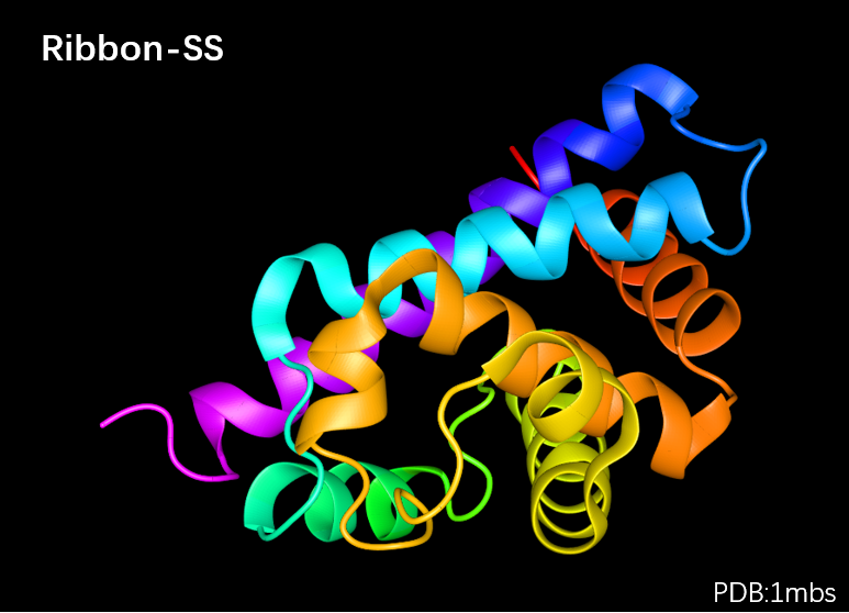
Menu → Main Structure → Ribbon-SS
The gallery of these styles is shown below.
| Line | Backbone | Sphere |
|---|---|---|
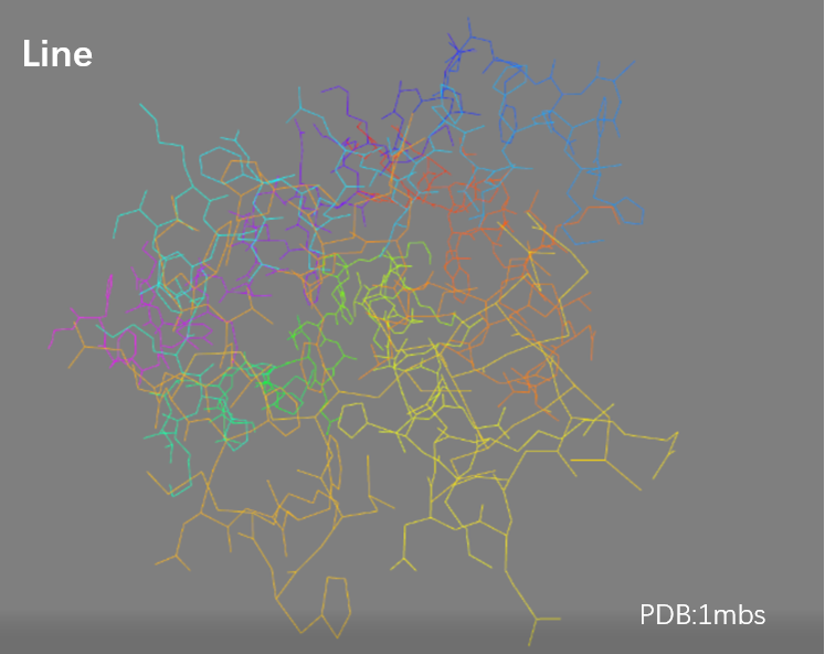 |  |  |
| Stick | Ball & Rod | Tube |
|---|---|---|
 |  |  |
| Ribbon-flat | Ribbon-ellipse | Ribbon-rectangle |
|---|---|---|
 |  |  |
| Ribbon-strip | Ribbon-railway | Ribbon-Secondary Structure |
|---|---|---|
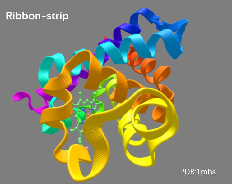 | 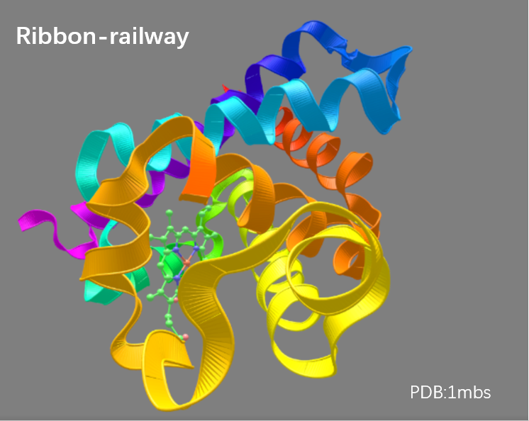 | 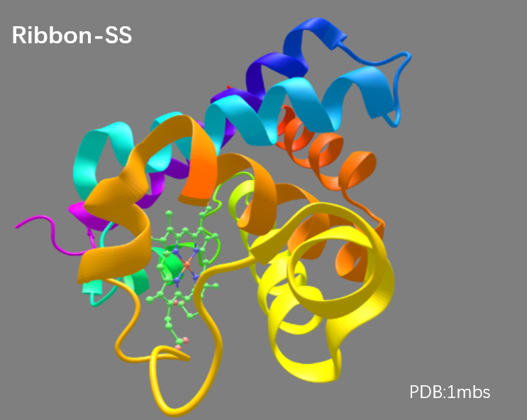 |
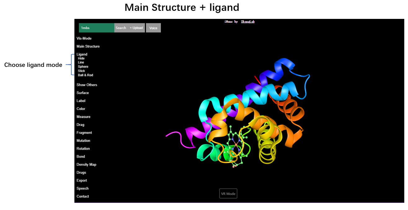
Ligand
Point the laser tip to "Ligand" in VR menu and press the trigger to choose the "ligand" option, and there will be 4 ligand representation styles in the submenu board: Line, Sphere, Stick, Ball & Rod.
Menu → Ligand → Line
Menu → Ligand → Sphere
Menu → Ligand → Stick
Menu → Ligand → Ball & Rod
The gallery of these styles is shown below.
| Line | Sphere | Stick | Ball & Rod |
|---|---|---|---|
 |  |  |  |
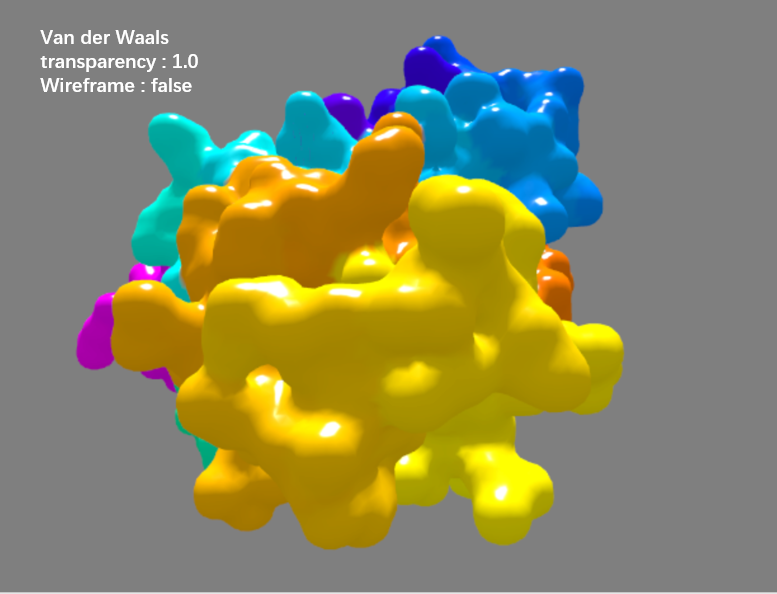
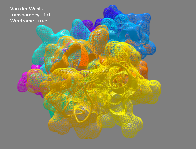
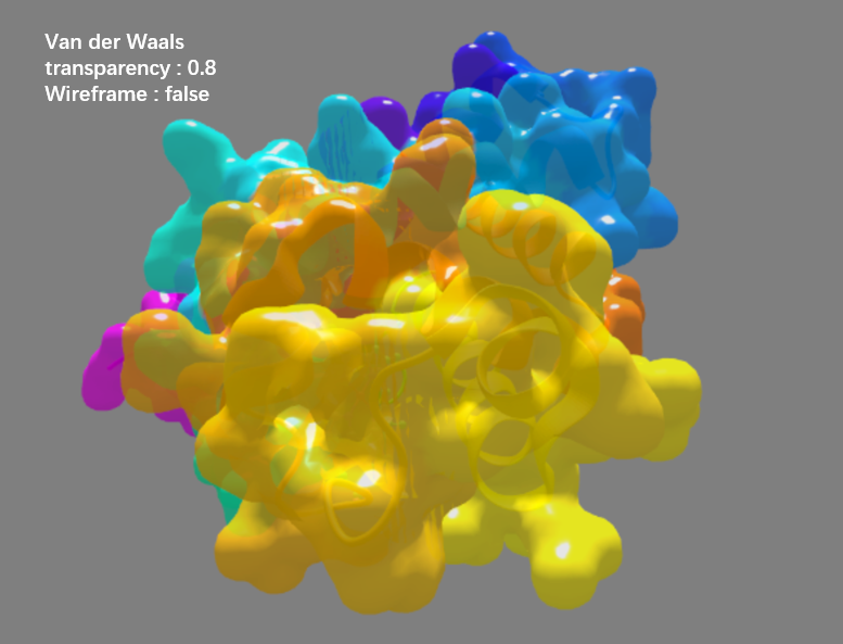
Surface
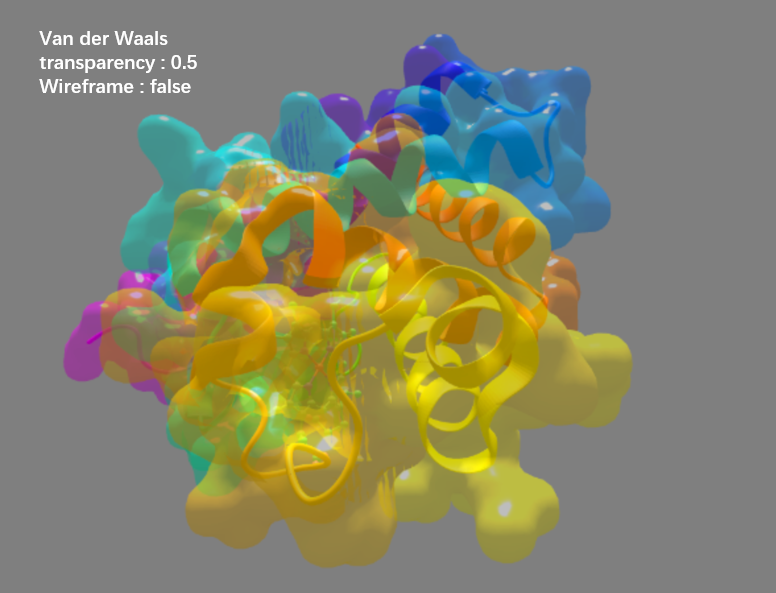
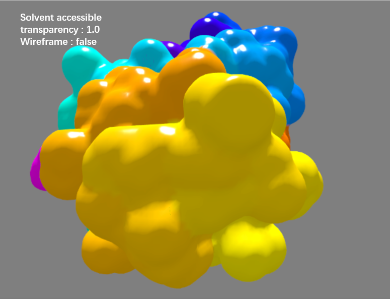
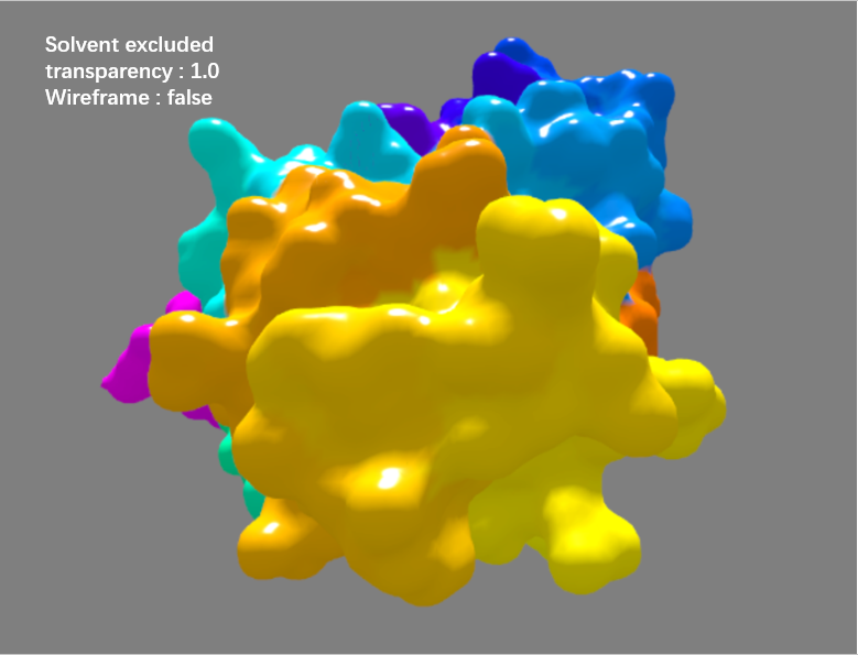
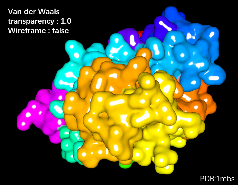
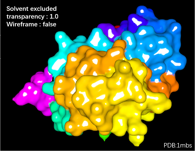
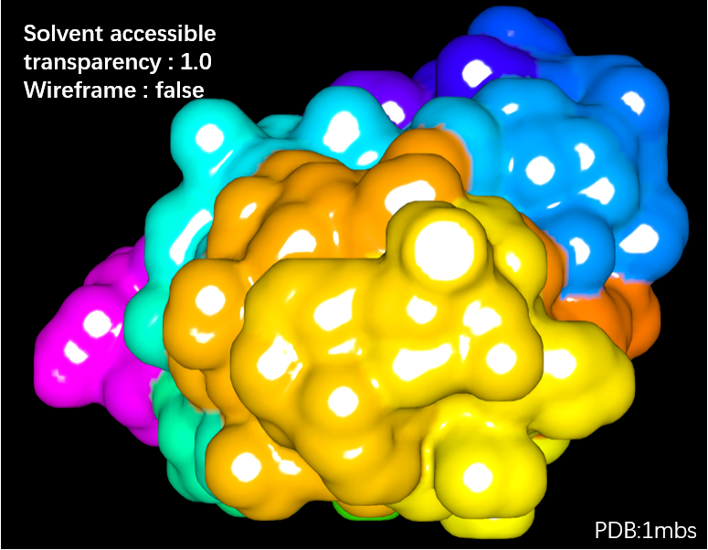
The "Surface" option includes Van der Waals, Solvent excluded, Solvent accessible and Molecular surface. The transparence of the structure surface can be adjusted by pointing the laser tip to Opacity 1.0-0.5 in submenu and pressing the trigger. In addition, by pointing the laser tip to Wireframe in submenu and pressing the trigger, the structure surface can be viewed as meshed surface.
Menu → Surface → Van der Waals surface
Menu → Surface → solvent accessible surface
Menu → Surface → solvent excluded surface
Menu → Surface → molecular surface
The gallery of these styles is shown below.
| Van der Waals surface | Van der Waals surface with Wireframesolvent accessible surface | Van der Waals surface with transparency(0.8) |
|---|---|---|
 |  |  |
| Van der Waals surface with transparency(0.5) | solvent accessible surface | solvent excluded surface |
|---|---|---|
 |  |  |
Color Scheme
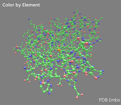
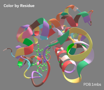
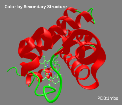
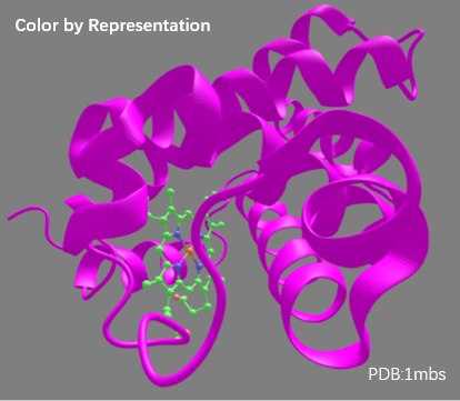
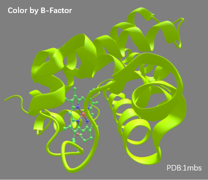
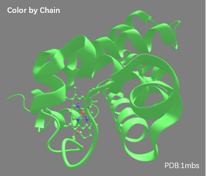
By choosing options in "Color" submenu in VR menu, the structure can be variedly colored according to 10 different schemes, such Element, Residue ,Secondary structure,Representation, Chain, B-factor, Spectrum, Chain Spectrum, Hydrophobicity, Sequence Conservation.
Menu → Color → Element
Menu → Color → Residue
Menu → Color → Secondary Structure
Menu → Color → Representation
Menu → Color → Chain
Menu → Color → B-factor
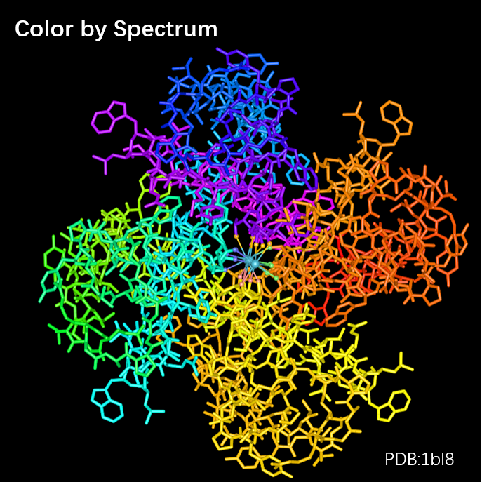
Menu → Color → Spectrum
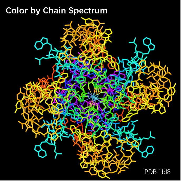
Menu → Color → Chain Spectrum
Menu → Color → Hydrophobicity
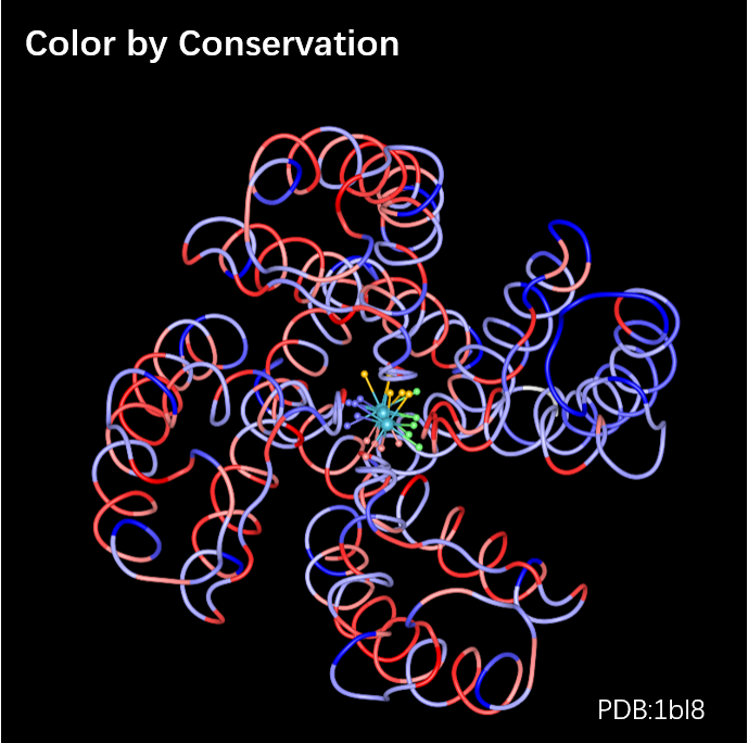
Menu → Color → Conservation

The gallery of these styles is shown below.
| Color by Element | Color by Residue | Color by Secondary Structure |
|---|---|---|
 |  |  |
| Color by Representation | Color by B-Factor | Color by Spectrum |
|---|---|---|
 |  | 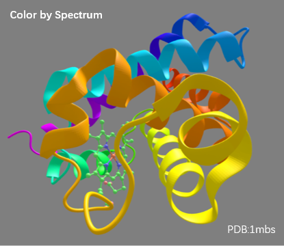 |
| Color by Chain Spectrum | Color by Hydrophobicity | Color by Conservation |
|---|---|---|
 | 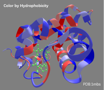 | 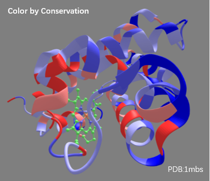 |
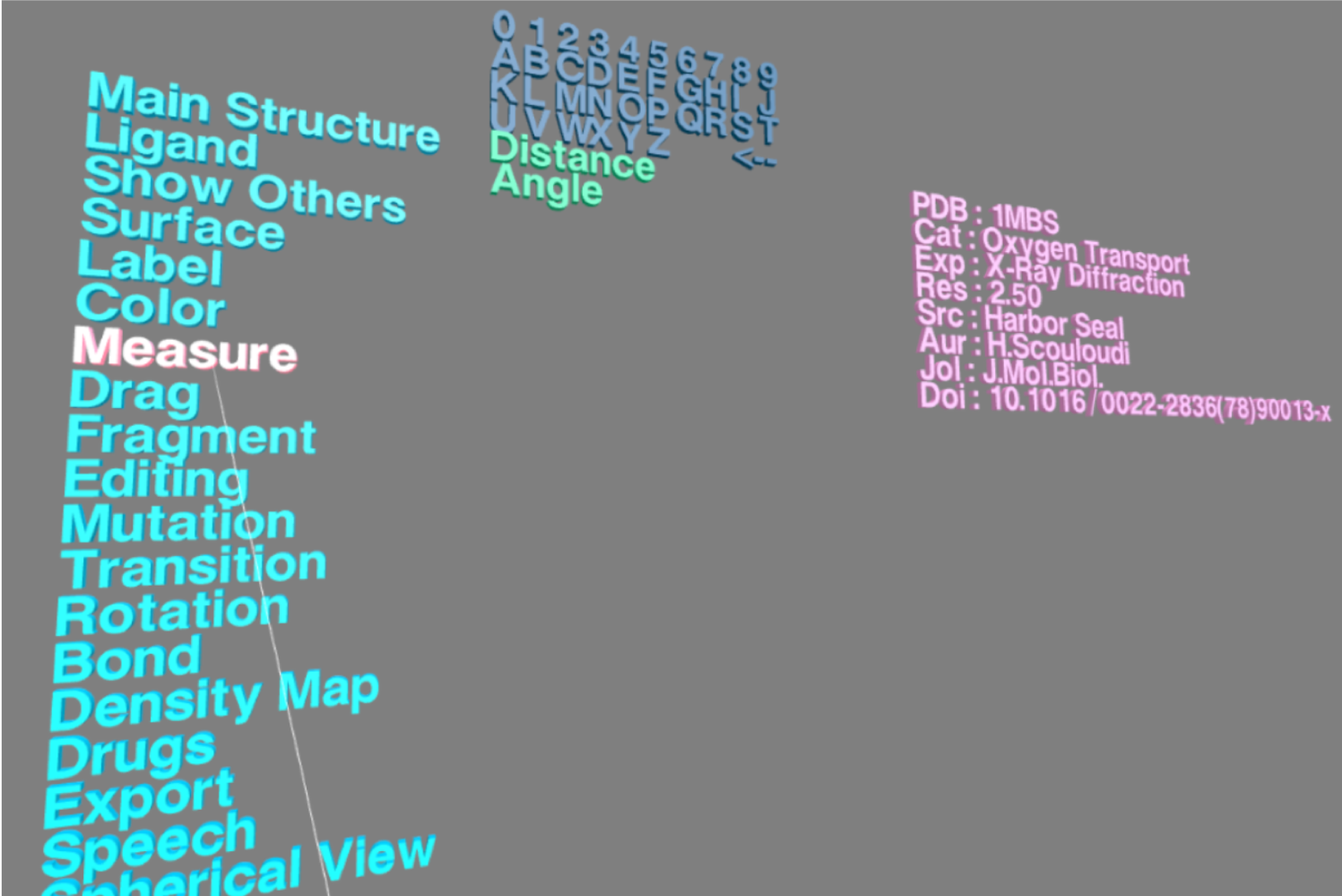
Measurement (V)
In VR menu, choose "Measure" and there will be Distance and Angle appearing in the submenu.
Point the laser tip to Distance option and press the trigger, the VR Scene containing the structure will appear. Point the laser tip to a residue and press the trigger to select it; then point the laser tip to another residue and press the trigger to select the second one. The distance between these two residues will be automatically presented.
Point the laser tip to Angle option and press the trigger, and then select three residues in the VR Scene. The angle among them will be automatically presented.
Menu → Measure → Distance
Menu → Measure → Angle
Here is an example video for computing distance in VR.
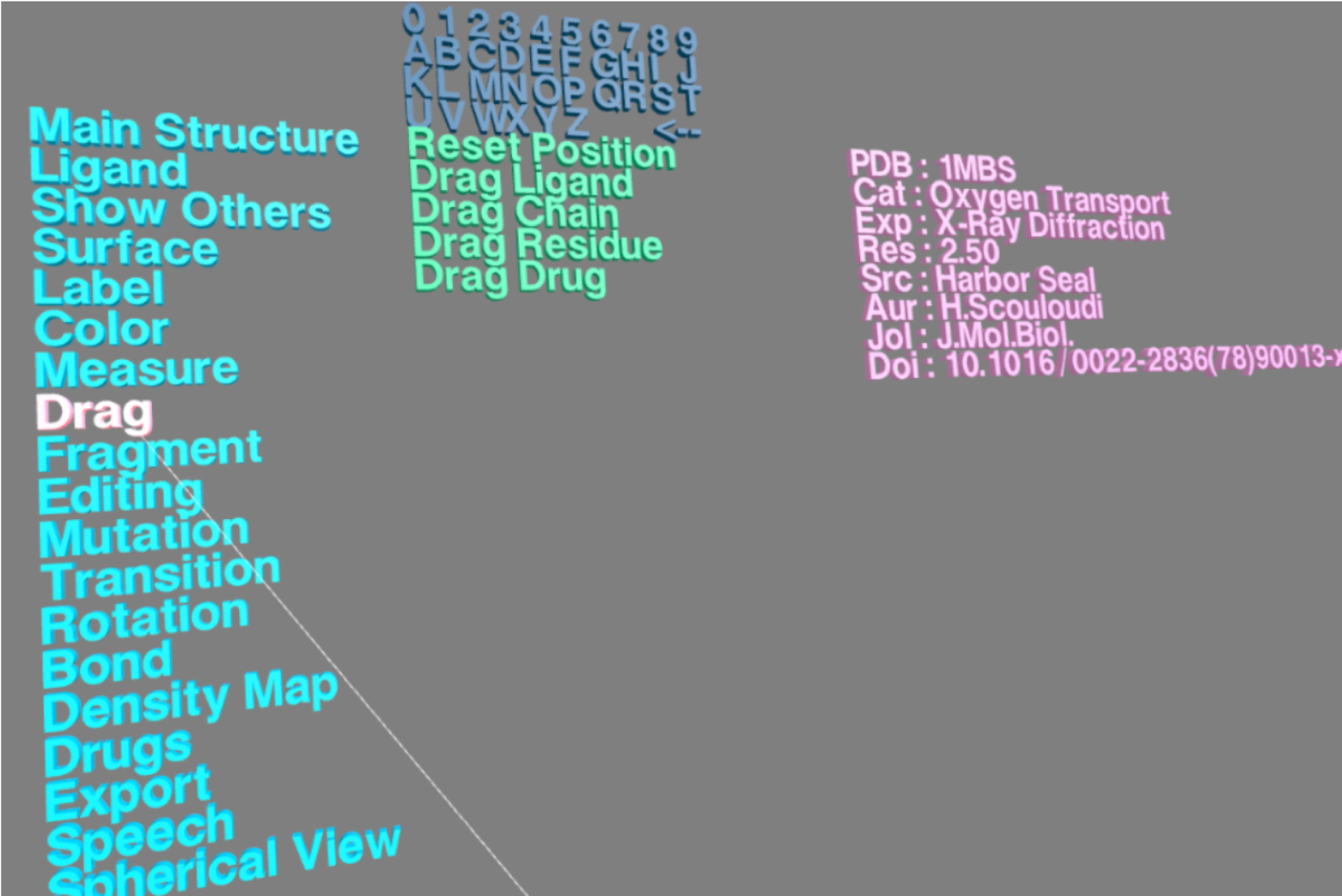
Drag (V)
"Drag" option is designed for dragging specific elements in the structure. By selecting Drag Chain, Drag Ligand, Drag Residue or Drag Drug, you can drag the individual chain, ligand, residue or drug of the structure in VR Scene.
Here is an example video for dragging specific elements in VR.
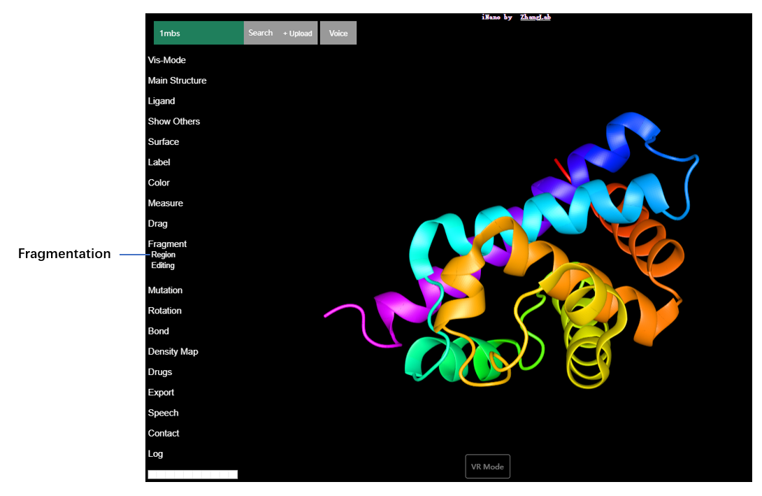
Fragmentation (V)
You can highlight specific fragments by presenting them with varied representation styles in the VR Scene. Firstly, enter "Fragment" in VR menu, and 12 representation styles will appear in submenu. Then choose a target representation style, and the VR Scene containing the input structure will apear. Next pick two residues by using the laser tip and pressing the trigger, and you will see the highlighted fragments presented with the previously-chosen representation style in VR scene.
Menu → Fragment
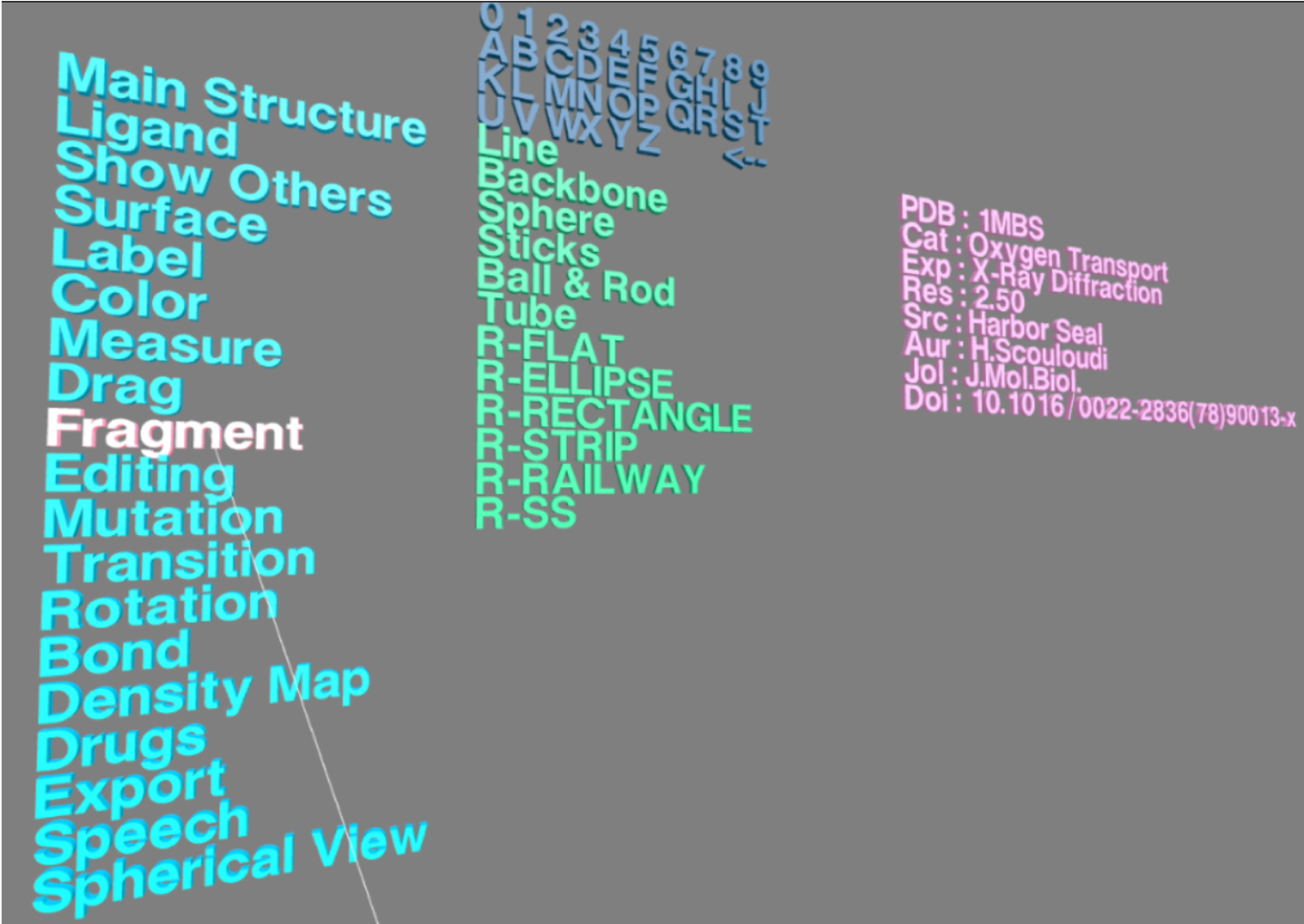
A selected fragment is highlighted with Ball&Rod style.
Here is an example video for fragments visualization.
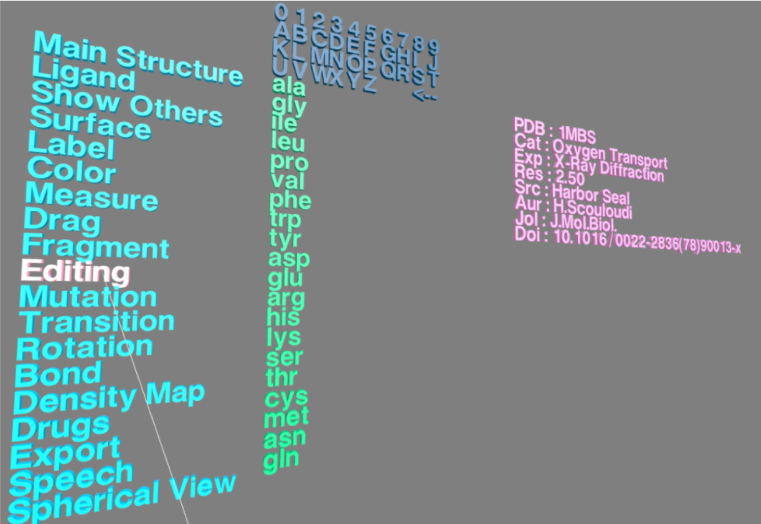
Structure Editing (V)
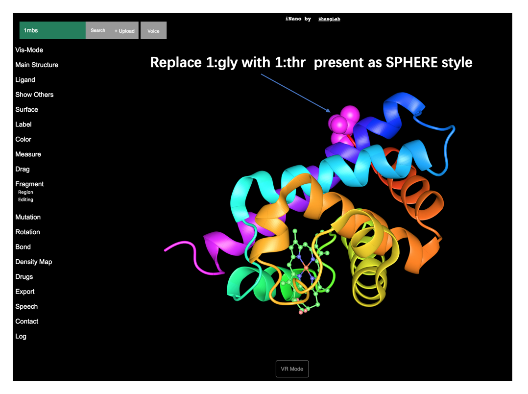
"Editing" provides a method to replace one amino acid with another one. Enter "Editing" in VR menu, and 20 types of amino acids will appear in the submenu. Choose the target type residue in the submenu by pointing the laser tip to it and pressing the trigger. Then pick the residue that you want to replace in the structure in VR Scene by pointing the laser tip to it and pressing the trigger, and you will see the picked residue is replaced by the target residue.
Menu → Editing
The edited structure can be exported as a new PDB file by using Export PDB in "Export" option in VR menu.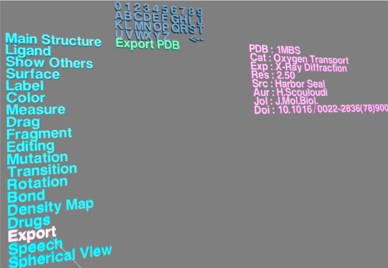
Here is an example video for structure editing in VR Scene.
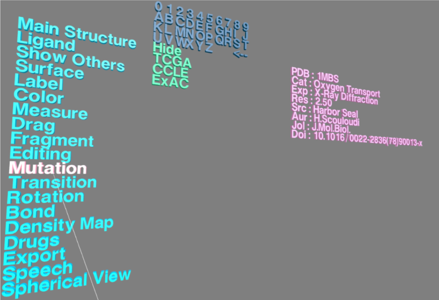
Genome Variations (V)
"Mutation" option in VR menu provides a handy way to view genomic variation sites in the target structure by connecting to TCGA, CCLE and ExAc databases. By choosing "Mutation" in VR menu, TCGA, CCLE and ExAC will apear in submenu board. And you can view genomic mutation sites (highlighted by twinkling balls) in the loaded structure by selecting these databases.
Menu → Mutation → TCGA
Menu → Mutation → CCLE
Menu → Mutation → ExAc
Here is an example video for genomic variations visualization. Try it
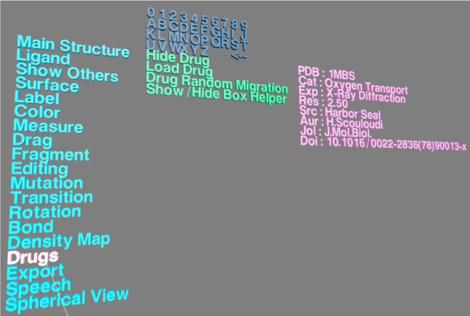
Interactive Docking (V)
In "Drugs" option in VR menu, choose Load Drug, and the structure-related drug (from DrugBank databse) will be loaded into the VR Scene. Press Menu button in the controller, and go back to VR menu. And choose Drug Random Migration to interactively perform drug docking in VR Scene.
Menu → Drugs → Load Drugs → docking
An example video for interactive docking in VR mode. Try it
Speech Recognition
VRmol provides speech recognition option, enabling users to operate the structure by speaking commands (the commands in English are listed below). Chinese commands can be accessed here.
| Category | Command |
| Visualization Mode | go to NonVR mode |
| go to virtual reality mode | |
| go to travel mode | |
| Main structure style | change to dot style |
| change to line style | |
| change to backbone style | |
| change to sphere style | |
| change to stick style | |
| change to ball and rod style | |
| change to tube style | |
| change to second structure style | |
| hide main structure | |
| show main structure | |
| Ligand structure style | change Ligand to line style |
| change Ligand to sphere style | |
| change Ligand to stick style | |
| change Ligand to ball and rod style | |
| hide ligand structure | |
| show ligand structure | |
| Show Other | show water |
| hide water | |
| show hydrogen bond | |
| hide hydrogen bond | |
| show mutation | |
| hide mutation | |
| Surface | show the surface of main structure |
| show transparency | |
| show mesh | |
| hide the surface of main structure | |
| Color | color by element |
| color by residue | |
| color by second structure | |
| color by chain | |
| color by b-factor | |
| color by spectrum | |
| color by hydrophobicity | |
| color by conservation | |
| Interaction | open dragging function |
| open labelling function | |
| rotation | |
| rotation by x | |
| rotation by y | |
| rotation by z | |
| clockwise rotation | |
| anticlockwise rotation | |
| move | |
| move along x | |
| move along y | |
| move along z | |
| move forward | |
| move backward |
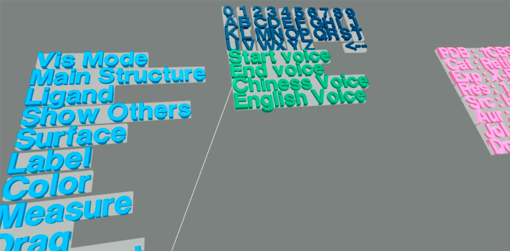
In VR menu, select "Speech" options. And then follow these steps:
- Choose a language by pointing the laser tip to Chinese Voice or English Voice and pressing the trigger. The default language is English.
- Point Voice in VR menu, and press down the trigger.
- Hold on pressing the trigger untill you finish speaking the command. The voice will be recorded and automatically uploaded to the server for recognization.
- VRmol will operate the structure according to the recognized command.
Menu → Speech → Voice
Here is an example video of speech-recognition.
NonVR Mode
Getting Started
Mouse Usage
Operation on the structure is controlled by mouse.
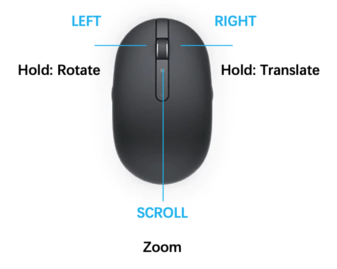
| Operation | Mouse Usage |
|---|---|
| Rotate | Press & hold LEFT mouse button, then move. |
| Translate | Press & hold RIGHT mouse button, then move. |
| Zoom | Scroll UP to zoom in; scroll DOWN to zoom out. |
Loading Structure
Users can access VRmol at https://vrmol.net through any WebGL supported browsers. Then type a PDB code (like 1MBS) in the textbox and then press enter button. A structure will appear in the user interface. In addition, VRmol also surpports loading structure by inputing a remote URL (such as https://files.rcsb.org/view/1mbs.pdb) or uploading a local structural file.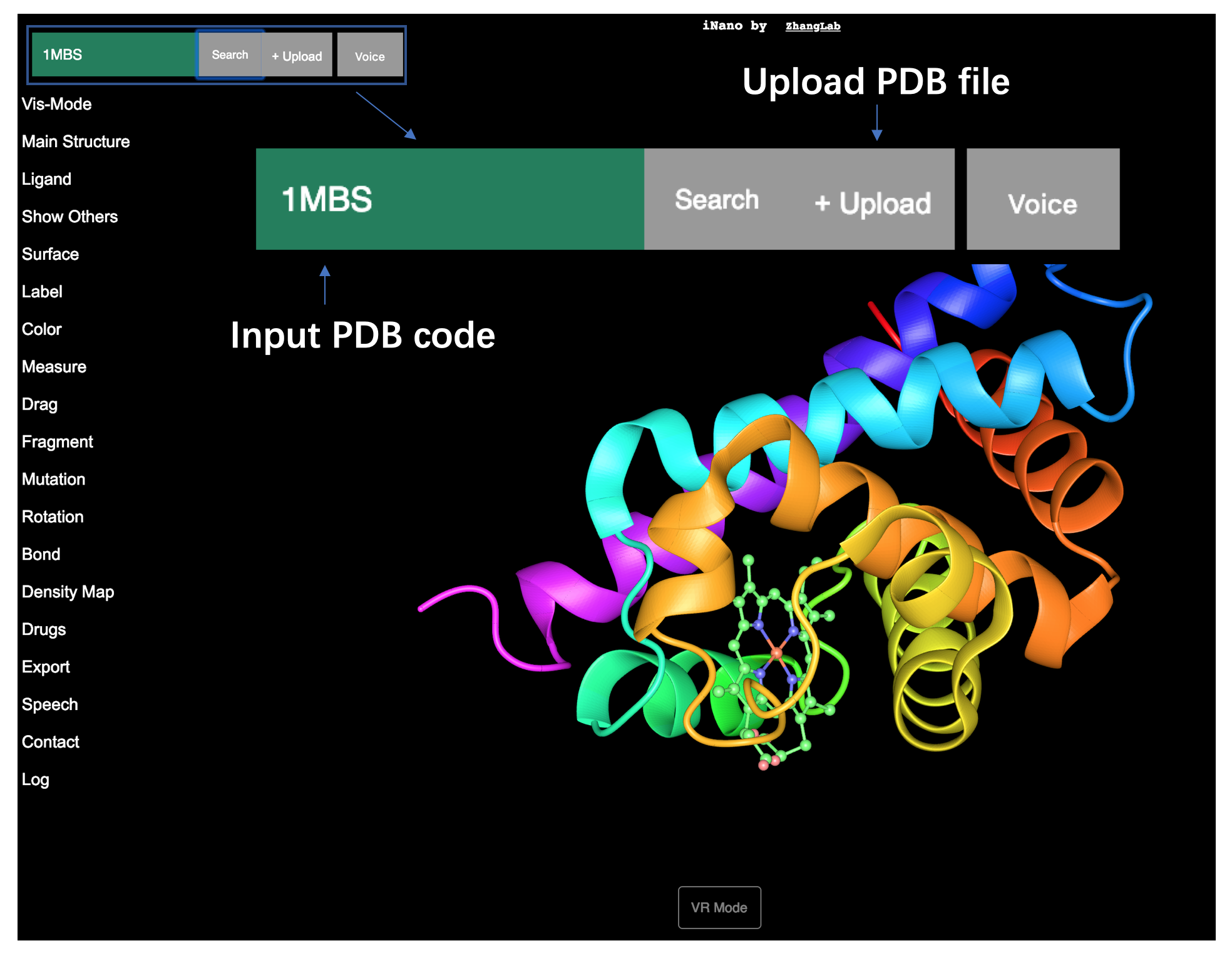
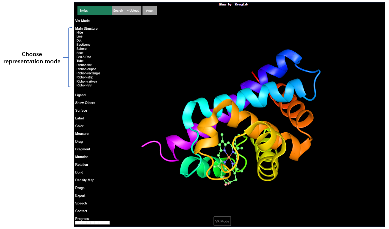
Main Structure
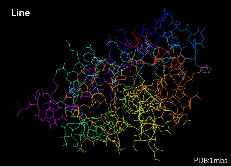
By clicking "Main Structure" option in the interface, you can view 12 different representation styles for "Main Structure", including Line, Backbone, Sphere, Stick, Ball & Rod, Tube, Ribbon-flat, Ribbon-ellipse, Ribbon-rectangle, Ribbon-strip, Ribbon-railway, Ribbon-SS.
Main Structure → Line
Main Structure → Backbone
Main Structure → Sphere
Main Structure → Stick
Main Structure → Ball & Rod
Main Structure → Tube
Main Structure → Ribbon-flat
Main Structure → Ribbon-ellipse
Main Structure → Ribbon-rectangle
Main Structure → Ribbon-ellipse
Main Structure → Ribbon-strip
Main Structure → Ribbon-railway
Main Structure → Ribbon-Secondary Structure
The gallery of the styles is shown below.
| Line | Backbone | Sphere |
|---|---|---|
 |  |  |
| Stick | Ball & Rod | Tube |
|---|---|---|
 |  |  |
| Ribbon-flat | Ribbon-ellipse | Ribbon-rectangle |
|---|---|---|
 |  |  |
| Ribbon-strip | Ribbon-railway | Ribbon-Secondary Structure |
|---|---|---|
 |  |  |
Ligand
By clicking "Ligand", you can select ligand representation styles.
You can view the ligand in four representation styles by clicking these affiliated options, including Line, Sphere, Stick, Ball & Rod .
Ligand → Line
Ligand → Sphere
Ligand → Stick
Ligand → Ball & Rod
| Line | Sphere | Stick | Ball & Rod |
|---|---|---|---|
 |  |  |  |
Surface
The "Surface" option includes Van der Waals, Solvent excluded, Solvent accessible and Molecular surface. The transparency of structure surface can be adjusted by clicking 1.0-0.5 in the affiliated options. In addition, by selecting Wireframe, the structure surface can be viewed as meshed surface.
Surface → Van der Waals surface
Surface → Solvent accessible surface
Surface → Solvent excluded surface
Surface → Molecular surface
Surface → 1.0 (Transparency)
Surface → 0.8 (Transparency)
Surface → Wireframe
The gallery of the surface representation styles is shown below.
| Van der Waals surface | Solvent excluded surface | Solvent accessible surface |
|---|---|---|
 |  |  |
| Molecular surface | Van der Waals surface with Wireframe | Van der Waals surface with transparency(0.8) |
|---|---|---|
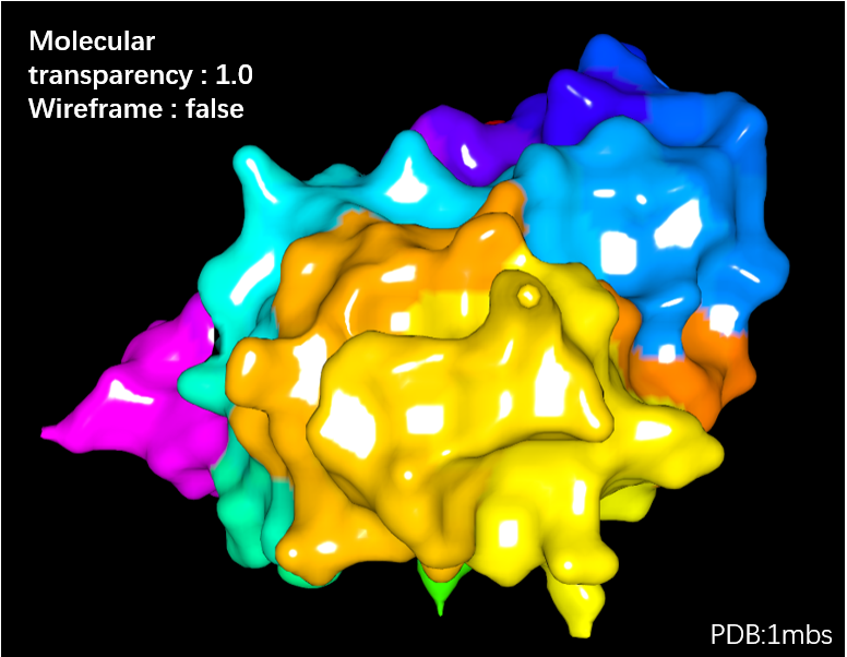 | 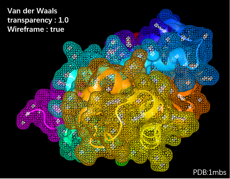 | 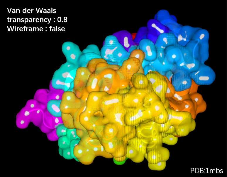 |
Color Scheme
By choosing options in "Color", the structure can be variedly colored according to 10 different schemes, such as Element, Residue ,Secondary structure,Representation, Chain, B-factor, Spectrum, Chain Spectrum, Hydrophobicity, Sequence Conservation.
Color → By Element
Color → By Residue
Color → By Second Structure
Color → By Representation
Color → By B-Factor
Color → By Spectrum
Color → By Chain Spectrum
Color → By Hydrophobicity
Color → By Conservation
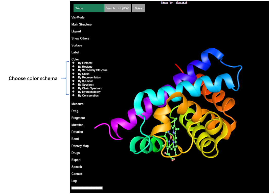
The gallery of the color schema is shown below.
| Color by Element | Color by Residue | Color by Second Structure |
|---|---|---|
 |  |  |
| Color by Representation | Color by B-Factor | Color by Spectrum |
|---|---|---|
 |  |  |
| Color by Chain Spectrum | Color by Hydrophobicity | Color by Conservation |
|---|---|---|
 |  |  |
Fragmentation (V)
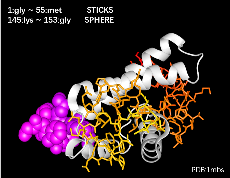
You can highlight specific fragments by presenting them with varied representation styles. Firstly, click "Fragment", and two affiliated options, Region and Editing, will appear.
Click the Region option, and the Fragment panel will appear as follows. The steps for representation of the selected fragments are labeled in the panel.
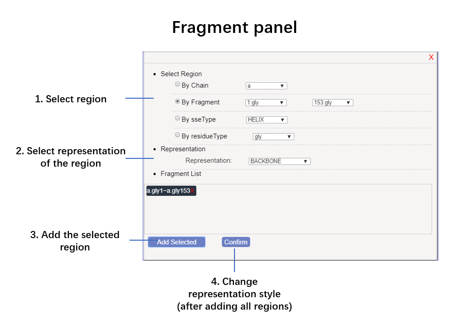
 Here is an example video for fragment regions.
Structure Editing (V)
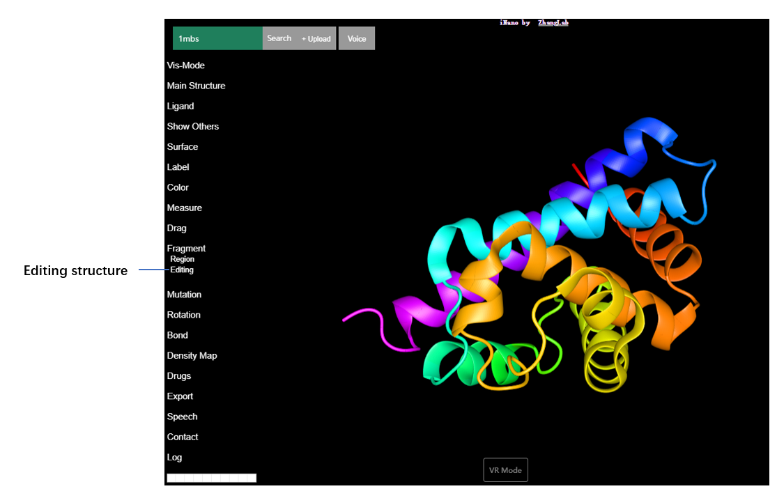
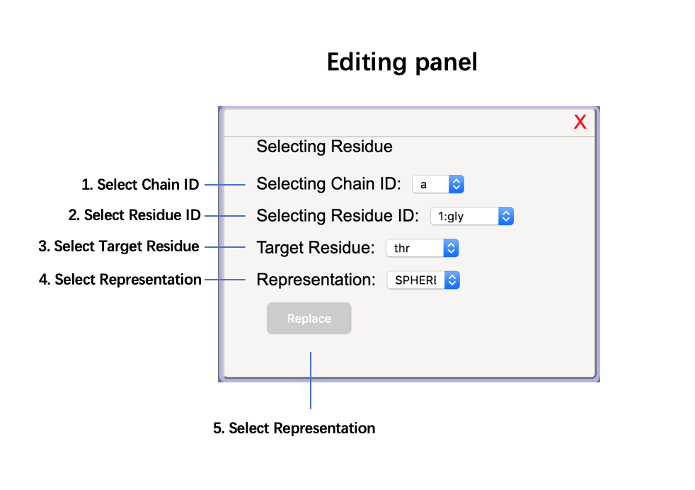
Structure Editing provides a method to replace one amino acid with another one. Click "Fragment", and then click its affiliated option Editing.
Here will appear an editing panel as follows. In this panel, you can select a specific chain by selecting its chain ID, and then select the residue that you want to replace. Next select the target residue and its representation style in the resulting structure. Finally, click Replace button in the panel, and you will see the original residue is replaced by the target residue highlighted by your selected representation style. Notably, the edited structure can be exported as a new PDB file by click "Export" option in the VRmol user interface.
Here is an example videos for structure editing in VRmol.
Genome Variations (V)
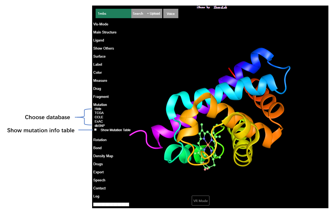
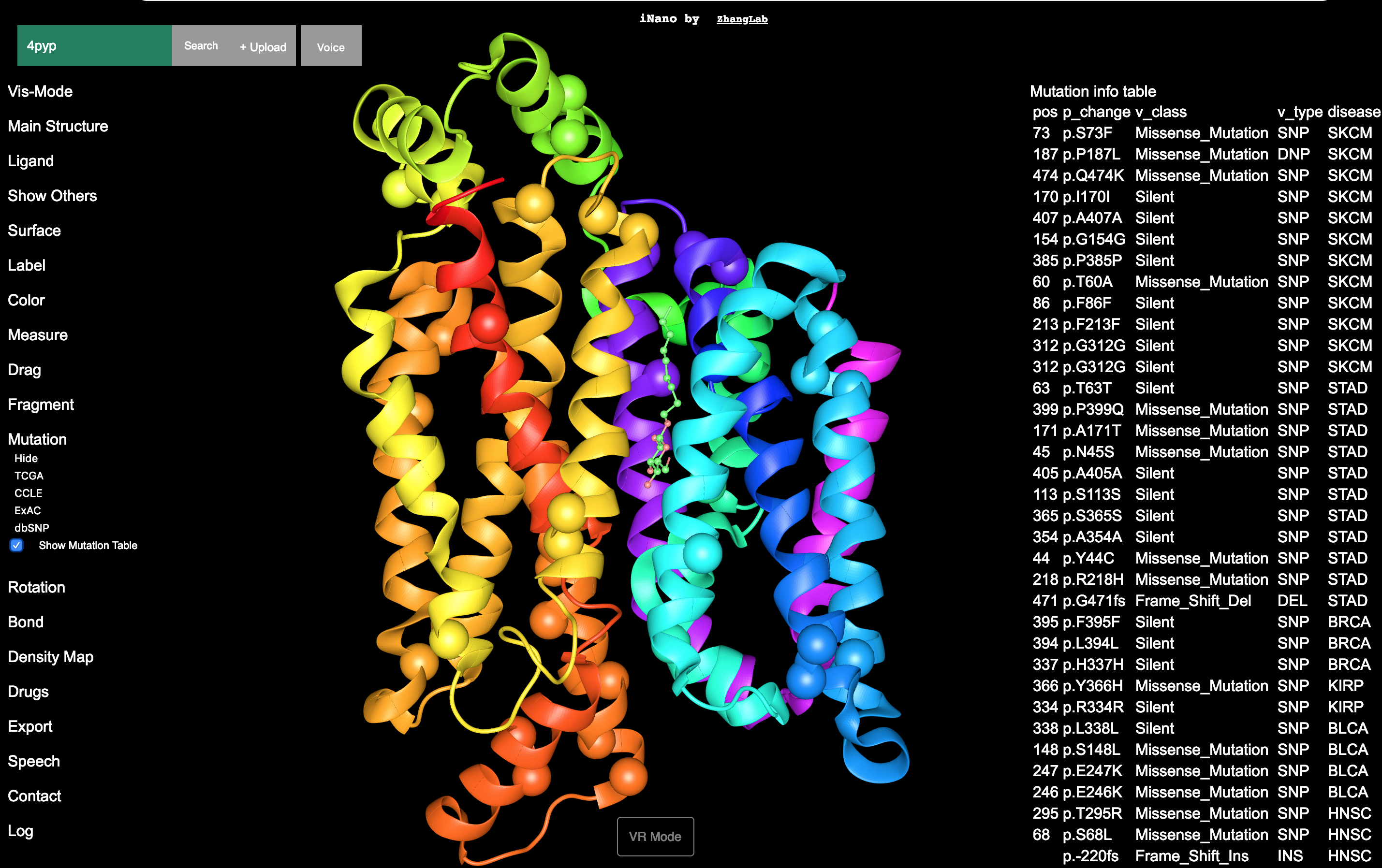
"Mutation" option provides a handy way to view genomic variation sites in the target structure by connecting to TCGA, CCLE, ExAc and dbSNP databases. By clicking "Mutation", TCGA, CCLE, ExAC and dbSNP will apear. And you can view genomic mutation sites (highlighted by twinkling balls) on the loaded structure by selecting these databases.
Mutation → TCGA
Mutation → CCLE
Mutation → ExAc
Mutation → dbSNP
Here is an example video for visualizing genomic variation. Try it
Interactive Docking (V)
"Drugs" option in VRmol provides a handy way for the structure-related drugs visualization and drug molecules docking analysis. In "Drugs" option, choose Load Drug, and the structure-related drug (from DrugBank databse) will be loaded into the user interface. And choose Drug Random Migration to perform drug docking. You can define the searching space in drug docking by clicking Show BoxHelper for getting more efficient and meaningful docking results. Once you click Show BoxHelper, there will appear a "Box Helper Limit" board in the upper right of the user interface, where searching space can be defined by inputing x, y and z coordinates.

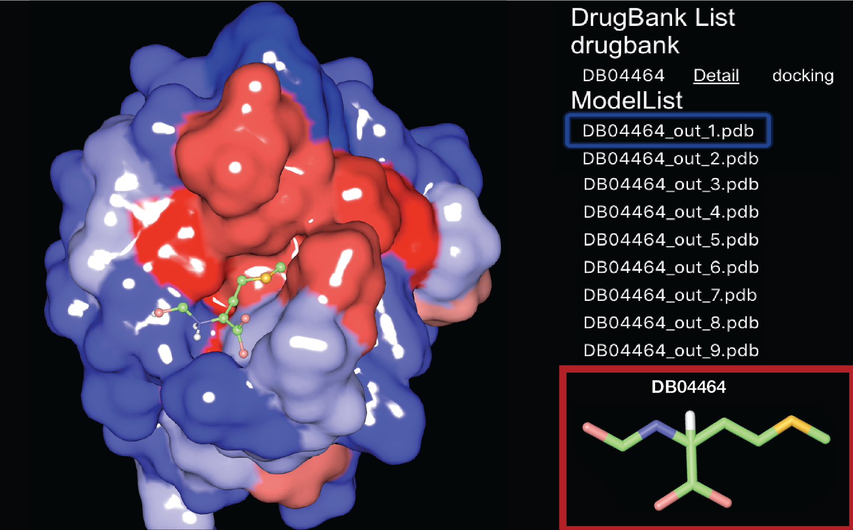
Here is an example video for drug docking. Try it
Density Map
If the input structure (PDB) has corresponding density map (like electron microscopy density map in EMDB), you can easily load this density map by clicking "Density Map" and then Load Density Map (as shown in the following figure). And the loaded density map can be visualized in several representation styles as listed in "Control panel for density map" in the following figure.
Density Map → Load Density Map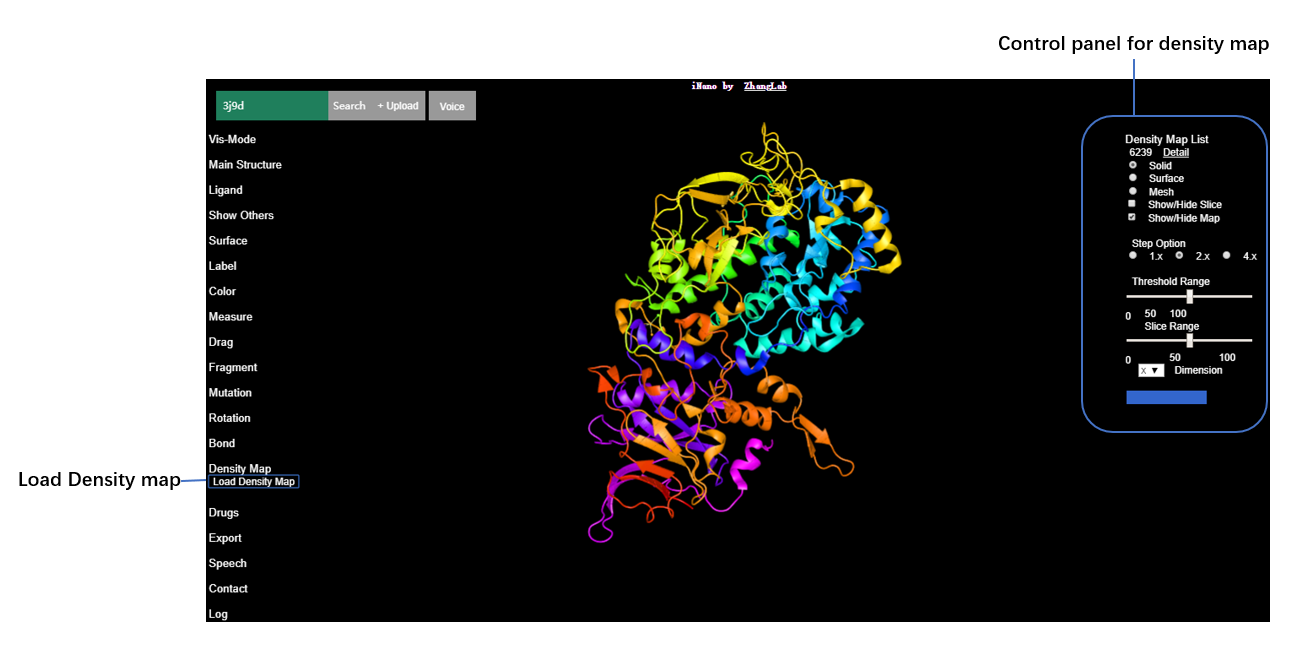
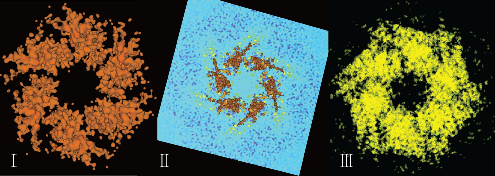
"Surface" (I), "Slice" (II) and "Solid" (III) representation styles of density map are shown below.
Notably, a local individual density map can also be directly uploaded by clicking "+Upload" button in the user interface.
Sharing by URL
URL Options
You can save a specific structural representation scene by saving operation options setting in VRmol as a URL, like https://vrmol.net/index.html?id=1dfb&panelShow=0&mainMode=7&showSurface=0&surfaceOpc=0.5&surfaceType=1&colorMode=602. And then you can load and share the scene by opening this previously-saved URL in the web browser or embedding it into your own website by using iframe, such as
1<iframe src="https://vrmol.net/index.html?id=1dfb&panelShow=0&mainMode=7&showSurface=1&surfaceOpc=0.5&surfaceType=1" />The options and their data types supported in VRmol are shown in the table below.
| Options | Data Type | Value | Comment |
|---|---|---|---|
| id | String | PDB code | 4 charactors |
| panelShow | Integer | Show:1,Hide:0 | Show menu panel or not |
| mainMode | Integer | LINE : 1, DOT : 2,BACKBONE : 3, SPHERE: 4,STICK : 5, BALL_AND_ROD : 6,TUBE : 7, RIBBON_FLAT:8, RIBBON_ELLIPSE:9, RIBBON_RECTANGLE:10, RIBBON_STRIP:11, RIBBON_RAILWAY:12, CARTOON_SSE:13,SURFACE:14 | Main Structure Representation |
| showSurface | Integer | Show:1,Hide:0 | Show surface panel or not |
| surfaceOpc | Float | 0~1 | Transparency of surface |
| surfaceType | Integer | Van der Waals surface:1, solvent excluded surface:2, solvent accessible surface:3, molecular surface:4 | Surface type |
| colorMode | Integer | Element:601, Residue:602, Secondary Structure:603, Chain:604, Representation:605, B-Factor:606", "Spectrum:607, Chain Spectrum:608, Hydrophobicity:609 | Color schemes |
| travel | Integer | Show:1,Hide:0 | Enter travel mode or not |
Examples for API
Present structure (PDB code:1MBS) with Ball & Rod style and hide the menu panel.
xxxxxxxxxx11<iframe src="https://vrmol.net/index.html?id=1mbs&panelShow=0&mainMode=6" />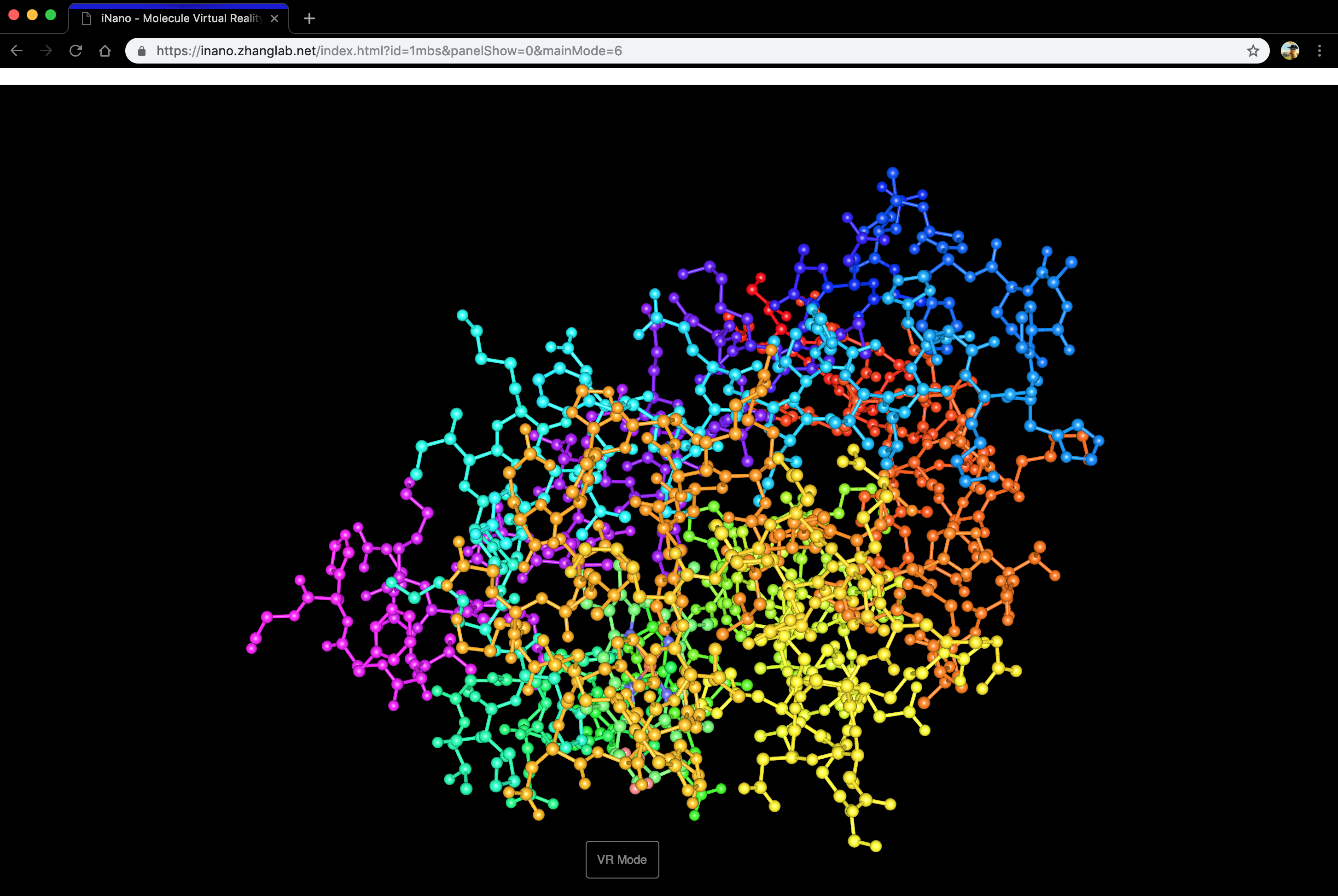
Present structure (PDB code:1DDB) with Tube style and show its Van der Waals surface with transparency (0.5), and hide the menu panel.
xxxxxxxxxx11<iframe src="https://vrmol.net/index.html?id=1dfb&panelShow=0&mainMode=7&showSurface=1&surfaceOpc=0.5&surfaceType=1" />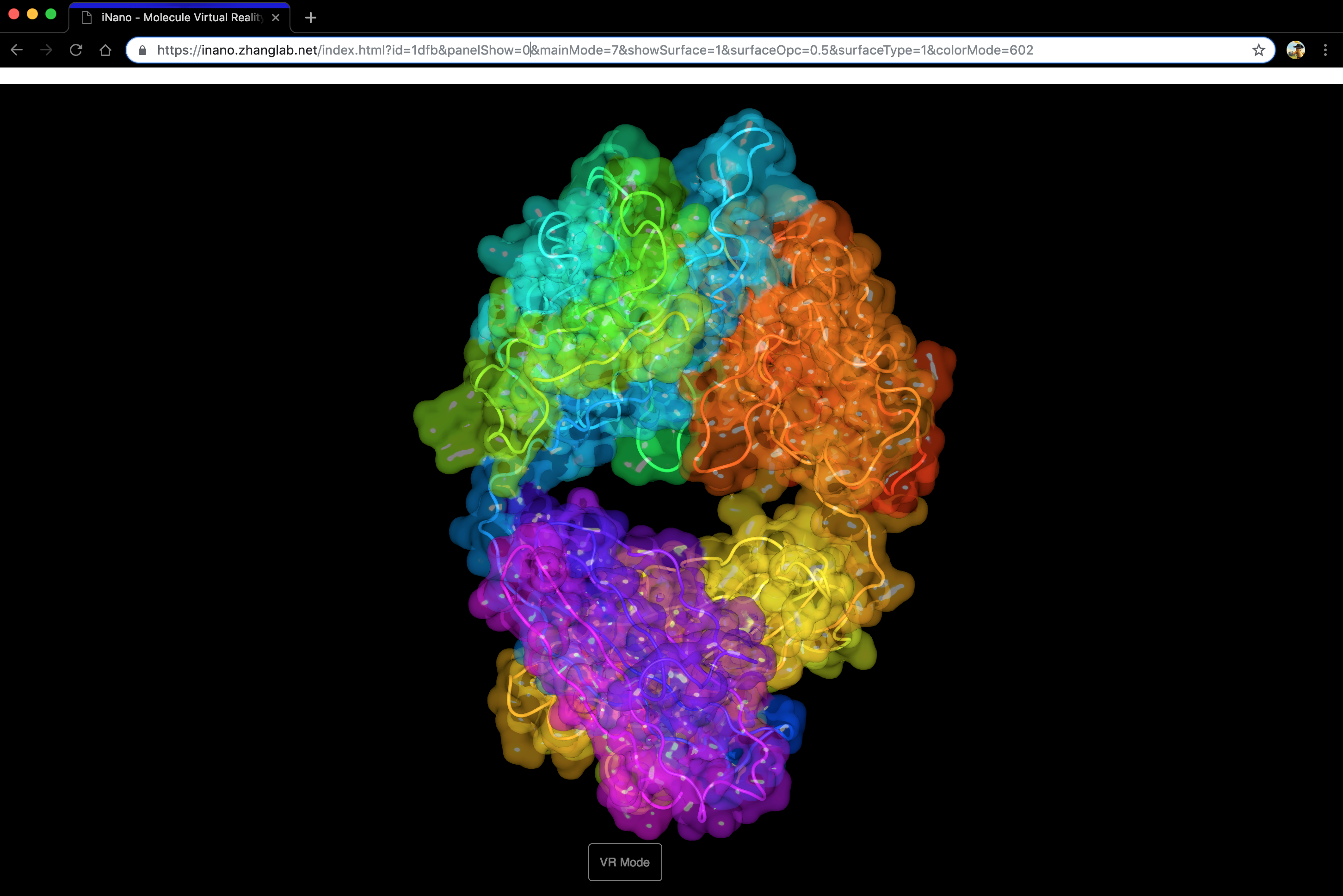
VRmol Service Access API
You can access VRmol Service (including drug auto-docking, genomic variations, speech recognition, sequence conservation visualization) by URL. Option settings are listed below. Once you request the service, VRmol will response and return with a json format string, which consists of data or warning information.
Genomic Variations
| Options | Data Type | Value | Comment |
|---|---|---|---|
| taskid | Integer | 10 | Request Genomic Variations for the provided structure |
| pdbid | String | PDB code | 4 charactors |
| ds | String | TCGA, CCLE, EXAC | Genomic Variations Database Name |
Example 1: https://vrmol.life.tsinghua.edu.cn/server/api.php?taskid=10&pdbid=1MBD&ds=tcga
xxxxxxxxxx41{2 "code": 0,3 "message": "no corresponding Uniprot-AC"4}Example 2: https://vrmol.life.tsinghua.edu.cn/server/api.php?taskid=10&pdbid=2YGD&ds=ccle
xxxxxxxxxx41{2 "code": 0,3 "message": "no mutation information in the current CCLE database."4}Example 3: https://vrmol.life.tsinghua.edu.cn/server/api.php?taskid=10&pdbid=2YGD&ds=tcga
xxxxxxxxxx991{2 "code": 1,3 "data": {4 "mutations": [5 {6 "id": "832951",7 "v_class": "Missense_Mutation",8 "v_type": "SNP",9 "g_change": "g.chr11:111779667C>G",10 "p_change": "p.E117Q",11 "disease": "UCEC",12 "pos": "117"13 },14 {15 "id": "832952",16 "v_class": "Missense_Mutation",17 "v_type": "SNP",18 "g_change": "g.chr11:111781056G>A",19 "p_change": "p.R107C",20 "disease": "UCEC",21 "pos": "107"22 },23 {24 "id": "832953",25 "v_class": "Silent",26 "v_type": "SNP",27 "g_change": "g.chr11:111781108G>T",28 "p_change": "p.L89L",29 "disease": "UCEC",30 "pos": "89"31 },32 {33 "id": "79882",34 "v_class": "Silent",35 "v_type": "SNP",36 "g_change": "g.chr11:111779560G>A",37 "p_change": "p.V152V",38 "disease": "SKCM",39 "pos": "152"40 },41 {42 "id": "118368",43 "v_class": "Missense_Mutation",44 "v_type": "SNP",45 "g_change": "g.chr11:111782377A>C",46 "p_change": "p.F24L",47 "disease": "SKCM",48 "pos": "24"49 },50 {51 "id": "390477",52 "v_class": "Silent",53 "v_type": "SNP",54 "g_change": "g.chr11:111782347C>T",55 "p_change": "p.E34E",56 "disease": "STAD",57 "pos": "34"58 },59 {60 "id": "646695",61 "v_class": "Missense_Mutation",62 "v_type": "SNP",63 "g_change": "g.chr11:111779603G>A",64 "p_change": "p.S138L",65 "disease": "LUAD",66 "pos": "138"67 }68 ],69 "chains": [70 "A",71 "B",72 "C",73 "D",74 "E",75 "F",76 "G",77 "H",78 "I",79 "J",80 "K",81 "L",82 "M",83 "N",84 "O",85 "P",86 "Q",87 "R",88 "S",89 "T",90 "U",91 "V",92 "W",93 "X"94 ],95 "pdbid": "2YGD",96 "dataset": "tcga"97 },98 "message": "success"99}
Sequence Conservation
| Options | Data Type | Value | Comment |
|---|---|---|---|
| taskid | Integer | 11 | Request Sequence Conservation for the provided structure |
| pdbid | String | PDB code | 4 charactors |
| chain | String | TCGA, CCLE, EXAC | Chain Name |
Example 1: https://vrmol.life.tsinghua.edu.cn/server/api.php?taskid=11&pdbid=3IVD&chain=A
xxxxxxxxxx301{2 code: 1,3 datas: [4 {5 pos: "1",6 seq: "M",7 resname: "",8 resid: "",9 chain: "",10 score: "0.013",11 color: "5",12 confinter: "-0.705,0.469",13 confintercolor: "7,4",14 msa: "8/301",15 resvar: "F,M,D,L"16 },17 {18 pos: "2",19 seq: "S",20 resname: "",21 resid: "",22 chain: "",23 score: "0.253",24 color: "4",25 confinter: "-0.355,0.687",26 confintercolor: "6,3",27 msa: "20/301",28 resvar: "A,S,T,D,K,G"29 },30
Drug data API
| Options | Data Type | Value | Comment |
|---|---|---|---|
| taskid | Integer | 12 | Request Related drugs for the provided structure |
| pdbid | String | PDB code | 4 charactors |
Example: http://vrmol.life.tsinghua.edu.cn/server/api.php?taskid=12&pdbid=2BR9
xxxxxxxxxx151{2 code: 1,3 data: [4 {5 id: "77",6 uniprotac: "P62258",7 bindingdb: "",8 chembl: "CHEMBL3329082;",9 swisslipids: "",10 guidetopharmacology: "",11 drugbank: "DB01780;"12 }13 ],14 message: "success"15}
EMDB
| Options | Data Type | Value | Comment |
|---|---|---|---|
| taskid | Integer | 13 | Request related electronic density map for the provided structure |
| pdbid | String | PDB code | 4 charactors |
Example: https://vrmol.life.tsinghua.edu.cn/server/api.php?taskid=13&pdbid=1MI6
xxxxxxxxxx111{2 code: 1,3 data: [4 "1006",5 "1007",6 "1008",7 "1009",8 "1010"9 ],10 message: "success"11}
AutoDock
| Options | Data Type | Value | Comment |
|---|---|---|---|
| taskid | Integer | 10 | Access AutoDock service |
| pdbid | String | PDB code | 4 charactors |
| smolid | String | drug name in DrugBank | drug name |
| x_c | Float | * | x value of center position of the docking area |
| y_c | Float | * | y value of center of the docking area |
| z_c | Float | * | z value of center of the docking area |
| x_s | Float | * | width value of the docking area |
| y_s | Float | * | height value of the docking area |
| z_s | Float | * | length value of the docking area |
xxxxxxxxxx101{2 jobid : "1mbs_DB04464_20181223200624_92",3 log : "http:\/\/vr.zhanglab.net\/server\/autodock\/jobs\/1mbs_DB04464_20181223200624_92\/log",4 pdbid : "1mbs",5 smolid : "DB04464",6 fullmodel : "DB04464_out.pdb",7 fullmodel_url : "http:\/\/vr.zhanglab.net\/server\/autodock\/jobs\/1mbs_DB04464_20181223200624_92\/DB04464_out.pdb",8 model_list": ["DB04464_out_1.pdb","DB04464_out_2.pdb","DB04464_out_3.pdb","DB04464_out_4.pdb","DB04464_out_5.pdb","DB04464_out_6.pdb","DB04464_out_7.pdb","DB04464_out_8.pdb","DB04464_out_9.pdb"],9 outdir : "http:\/\/vr.zhanglab.net\/server\/autodock\/jobs\/1mbs_DB04464_20181223200624_92"10}
Speech Recognition
For accessing Speech Recognition service, you only need to import https://vrmol.net/server/listen/recorder.js into your html code.
Example:
xxxxxxxxxx161<html>2 <meta http-equiv="Content-Type"content="text/html; charset=utf-8"/>3 <script src="jquery-3.2.1.min.js"></script>4 <script src="https://vrmol.net/server/listen/recorder.js"></script>5 <body>6 <div class="messages"></div>7 <button class="test" onmousedown="startRecording()" onmouseup="endRecording()">Voice</button>8 <br>9 <input name="language" value="Chinese" type="radio" id="Chinese" onclick="getlanguage()" checked="true">10 <label for="Chinese">For Chinese</label>11 <br>12 <input name="language" value="English" type="radio" id="English" onclick="getlanguage()">13 <label for="English">For English</label>14 <br>15 </body>16</html>Contacts
VRmol is developed and supported by ZhangLab and WangLab in Tsinghua University. Any question about VRmol, please contact xuk16@mails.tsinghua.edu.cn.